Breast Cancer,Illustration
Bildnummer 12036991
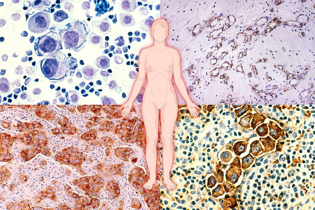
| Illustration of the female body superimposed on a composite of micrographs of breast cancer cells. Clockwise from top left: (1) Human metastatic breast cancer in the pleural fluid. Stained with HandE and magnified to 400x. (2) Cross section of infiltrating ductal carcinoma of the breast with a small foci of breast cancer cells in which cd34 antibody has stained blood vessels and basement membrane. Magnification x100. (3) Human metastatic breast cancer in the lymph nodes. Stained by immunocytochemical for epithelial membrane antigen. Magnified to 400x. (4) An infiltrating ductal carcinoma of human breast origin is seen invading the breast tissue. The cytoplasm of the tumour cells is stained brown with a monoclonal antibody,which recognizes a carcinoembryonic type antigen (CEA) found within the malignant cells. Magnification is 313x | |
| Lizenzart: | Lizenzpflichtig |
| Credit: | Science Photo Library / Martin, Mary |
| Bildgröße: | 5272 px × 3512 px |
| Modell-Rechte: | nicht erforderlich |
| Eigentums-Rechte: | nicht erforderlich |
| Restrictions: |
|
Preise für dieses Bild ab 15 €
Universitäten & Organisationen
(Informationsmaterial Digital, Informationsmaterial Print, Lehrmaterial Digital etc.)
ab 15 €
Redaktionell
(Bücher, Bücher: Sach- und Fachliteratur, Digitale Medien (redaktionell) etc.)
ab 30 €
Werbung
(Anzeigen, Aussenwerbung, Digitale Medien, Fernsehwerbung, Karten, Werbemittel, Zeitschriften etc.)
ab 55 €
Handelsprodukte
(bedruckte Textilie, Kalender, Postkarte, Grußkarte, Verpackung etc.)
ab 75 €
Pauschalpreise
Rechtepakete für die unbeschränkte Bildnutzung in Print oder Online
ab 495 €
Keywords
- Anatomie,
- anatomisch,
- Antigen,
- Brust,
- Brustkrebs,
- cea,
- Duktalkarzinom,
- Gewebe,
- Histologie,
- histologisch,
- Illustration,
- Karzinom,
- Krankheit,
- Krebs,
- krebsartig,
- Krebszelle,
- Lichtmikroskop,
- lichtmikroskopische Aufnahme,
- Lichtmikroskopische Aufnahmen,
- lms,
- Lymphknoten,
- maligne,
- Malignom,
- medizinisch,
- Metastase,
- Mikrofotografie,
- Mikrophotographie,
- Mikroskopie,
- Mikroskopische Aufnahmen,
- monoklonaler Antikörper,
- Pathologie,
- Querschnitt,
- Tumor,
- Verfärbung,
- Wissenschaft,
- Zeichnung,
- Zellen,
- zellular,
- Zytoplasma