Sinusitis with Empyema,MRI
Bildnummer 12036620
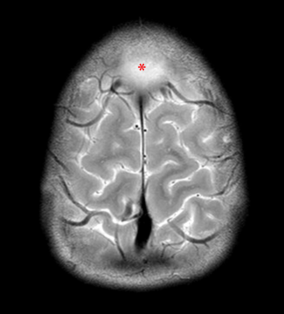
| This axial (cross-sectional),T2-weighted MR shows an extra-axial (outside the brain) collection of increased T2 signal (looks whiter than normal) in the frontal midline (asterisk - near the top of the image),compressing the superior sagital sinus. Further imaging revealed that this represents and area of edema and cerebritis (brain inflammation) complicating frontal sinusitis | |
| Lizenzart: | Lizenzpflichtig |
| Credit: | Science Photo Library / Living Art Enterprises |
| Bildgröße: | 4200 px × 4641 px |
| Modell-Rechte: | nicht erforderlich |
| Eigentums-Rechte: | nicht erforderlich |
| Restrictions: |
|
Preise für dieses Bild ab 15 €
Universitäten & Organisationen
(Informationsmaterial Digital, Informationsmaterial Print, Lehrmaterial Digital etc.)
ab 15 €
Redaktionell
(Bücher, Bücher: Sach- und Fachliteratur, Digitale Medien (redaktionell) etc.)
ab 30 €
Werbung
(Anzeigen, Aussenwerbung, Digitale Medien, Fernsehwerbung, Karten, Werbemittel, Zeitschriften etc.)
ab 55 €
Handelsprodukte
(bedruckte Textilie, Kalender, Postkarte, Grußkarte, Verpackung etc.)
ab 75 €
Pauschalpreise
Rechtepakete für die unbeschränkte Bildnutzung in Print oder Online
ab 495 €
Keywords
- abnormal,
- Anomalie,
- Bildgebung,
- cerebral,
- Diagnose,
- diagnostische Bildgebung,
- entzündet,
- Entzündung,
- Gehirn,
- geschwollen,
- Hirnhaut-,
- Infektion,
- Kondition,
- Kopf,
- Krankheit,
- Medizin,
- medizinisch,
- medizinische Bildgebung,
- Meningitis,
- MRI,
- Nebenhöhlen,
- Nebenhöhlenentzündung,
- Pathologie,
- Schwellung,
- Sinus,
- Störung,
- subdural,
- ungesund,
- Wissenschaft