Sinusitis with Empyema,CT Scan
Bildnummer 12036612
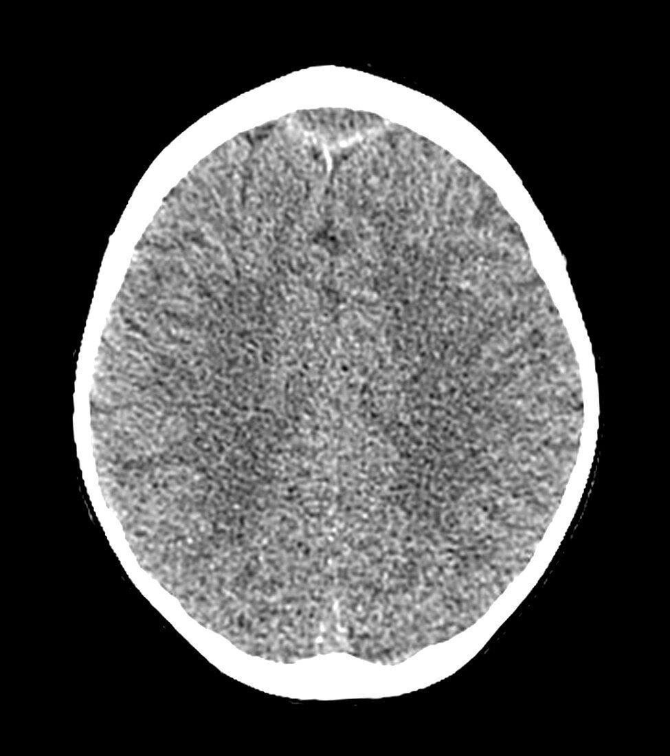
| This axial (cross-sectional) CT image shows an area of hypodensity (darker than normal) just anterior (towards the top of the image) to the superior sagital sinus. Further imaging revealed that this represents and area of edema/cerebritis (brain inflammation),subdural and epidural empyemas (abscess collections) complicating frontal sinusitis | |
| Lizenzart: | Lizenzpflichtig |
| Credit: | Science Photo Library / Living Art Enterprises |
| Bildgröße: | 4200 px × 4740 px |
| Modell-Rechte: | nicht erforderlich |
| Eigentums-Rechte: | nicht erforderlich |
| Restrictions: |
|
Preise für dieses Bild ab 15 €
Universitäten & Organisationen
(Informationsmaterial Digital, Informationsmaterial Print, Lehrmaterial Digital etc.)
ab 15 €
Redaktionell
(Bücher, Bücher: Sach- und Fachliteratur, Digitale Medien (redaktionell) etc.)
ab 30 €
Werbung
(Anzeigen, Aussenwerbung, Digitale Medien, Fernsehwerbung, Karten, Werbemittel, Zeitschriften etc.)
ab 55 €
Handelsprodukte
(bedruckte Textilie, Kalender, Postkarte, Grußkarte, Verpackung etc.)
ab 75 €
Pauschalpreise
Rechtepakete für die unbeschränkte Bildnutzung in Print oder Online
ab 495 €
Keywords
- abnormal,
- Anomalie,
- Bildgebung,
- cerebral,
- Computertomographie,
- ct,
- CT-Scan,
- Diagnose,
- diagnostische Bildgebung,
- entzündet,
- Entzündung,
- Gehirn,
- geschwollen,
- Hirnhaut-,
- Infektion,
- Kondition,
- Kopf,
- Krankheit,
- Medizin,
- medizinisch,
- medizinische Bildgebung,
- Meningitis,
- Nebenhöhlen,
- Nebenhöhlenentzündung,
- Neuroimaging,
- Pathologie,
- Schwellung,
- Sinus,
- Störung,
- subdural,
- ungesund,
- Wissenschaft