Sinusitis Complicated by Empyema,CT Scan
Bildnummer 12036587
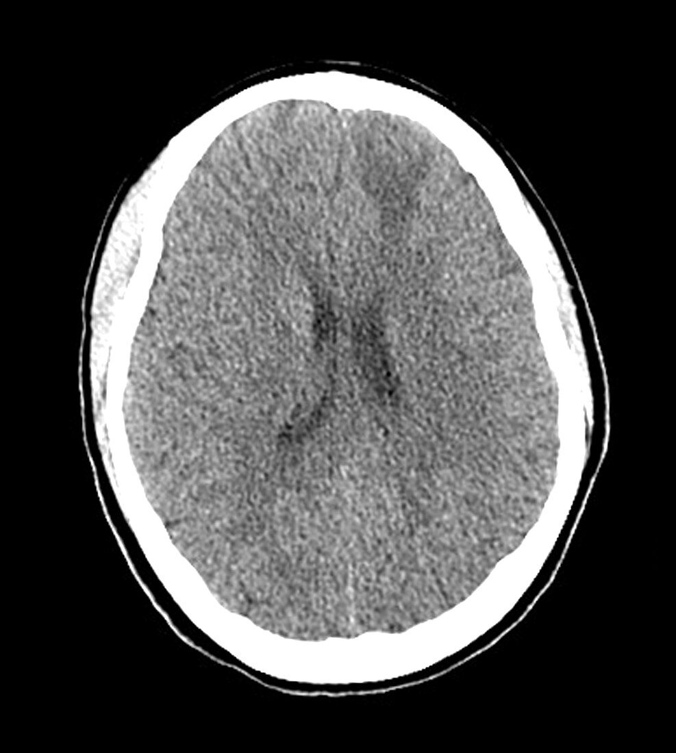
| This axial (cross-sectional) CT image shows an area of hypodensity (darker than normal) in the frontal lobe on viewer's right. Further imaging revealed that this represents and area of edema and cerebritis (brain inflammation) complicating frontal sinusitis | |
| Lizenzart: | Lizenzpflichtig |
| Credit: | Science Photo Library / Living Art Enterprises |
| Bildgröße: | 4200 px × 4679 px |
| Modell-Rechte: | nicht erforderlich |
| Eigentums-Rechte: | nicht erforderlich |
| Restrictions: |
|
Preise für dieses Bild ab 15 €
Universitäten & Organisationen
(Informationsmaterial Digital, Informationsmaterial Print, Lehrmaterial Digital etc.)
ab 15 €
Redaktionell
(Bücher, Bücher: Sach- und Fachliteratur, Digitale Medien (redaktionell) etc.)
ab 30 €
Werbung
(Anzeigen, Aussenwerbung, Digitale Medien, Fernsehwerbung, Karten, Werbemittel, Zeitschriften etc.)
ab 55 €
Handelsprodukte
(bedruckte Textilie, Kalender, Postkarte, Grußkarte, Verpackung etc.)
ab 75 €
Pauschalpreise
Rechtepakete für die unbeschränkte Bildnutzung in Print oder Online
ab 495 €
Keywords
- abnormal,
- Bildgebung,
- Computertomographie,
- ct,
- CT-Scan,
- Diagnose,
- diagnostische Bildgebung,
- entzündet,
- Entzündung,
- Gehirn,
- geschwollen,
- Hirnhaut-,
- Infektion,
- Kondition,
- Kopf,
- Krankheit,
- Medizin,
- medizinisch,
- medizinische Bildgebung,
- Meningitis,
- Nebenhöhlenentzündung,
- Neuroimaging,
- Pathologie,
- Schwellung,
- Störung,
- subdural,
- ungesund,
- Wissenschaft