Haemorrhagic Cerebral Infarct,MRI
Bildnummer 12036577
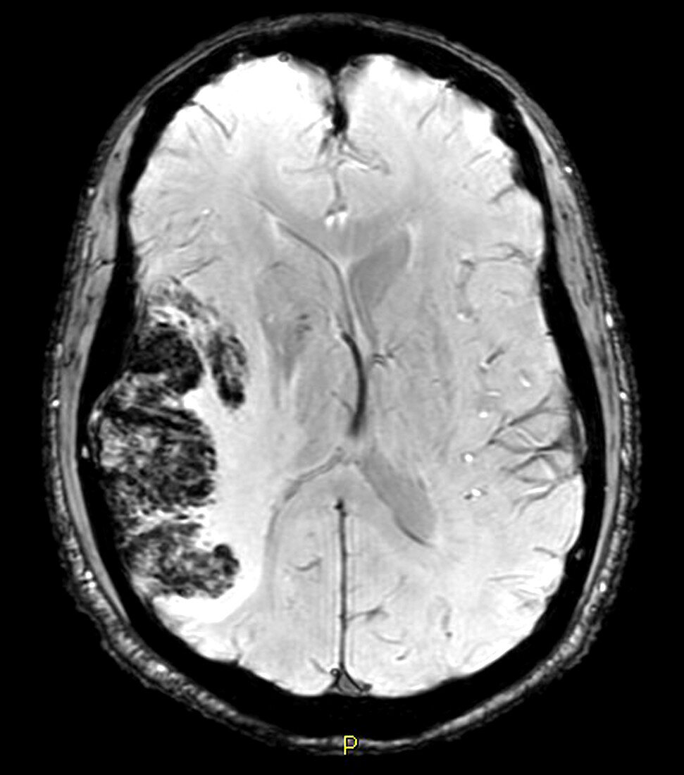
| This axial (cross-sectional) susceptibility weighted image (SWI) of the brain demonstrates a large area of abnormal increased signal (looks whiter than normal) in the middle cerebral artery vascular territory (on viewer's left) compatible with an acute infarct (stroke). There are also areas of darker signal along the lateral margin of this infarct which represent blood (hemorrhage). SWI is a new pulse sequence in MRI. This technique is exquisitely sensitive to the presence of blood and calcium and can be very abnormal even in the setting of normal routine MR images | |
| Lizenzart: | Lizenzpflichtig |
| Credit: | Science Photo Library / Living Art Enterprises |
| Bildgröße: | 4200 px × 4757 px |
| Modell-Rechte: | nicht erforderlich |
| Eigentums-Rechte: | nicht erforderlich |
| Restrictions: |
|
Preise für dieses Bild ab 15 €
Universitäten & Organisationen
(Informationsmaterial Digital, Informationsmaterial Print, Lehrmaterial Digital etc.)
ab 15 €
Redaktionell
(Bücher, Bücher: Sach- und Fachliteratur, Digitale Medien (redaktionell) etc.)
ab 30 €
Werbung
(Anzeigen, Aussenwerbung, Digitale Medien, Fernsehwerbung, Karten, Werbemittel, Zeitschriften etc.)
ab 55 €
Handelsprodukte
(bedruckte Textilie, Kalender, Postkarte, Grußkarte, Verpackung etc.)
ab 75 €
Pauschalpreise
Rechtepakete für die unbeschränkte Bildnutzung in Print oder Online
ab 495 €