Carotid Cavernous Sinus Fistula,MRI
Bildnummer 12036542
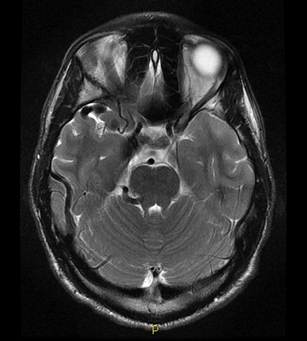
| This axial,T2-weighted MR image demonstrates pathologic enlargement of the cavernous sinus (on the viewer's left),in addition to abnormal,enlarged vascular flow voids (black) in the anterior middle cranial fossa. These changes reflect a post-traumatic carotid cavernous sinus fistula. These occur as a result of a traumatic tear of the internal carotid artery within the cavernous sinus,resulting in an arterial-venous shunt directly into the cavernous sinus and its venous outflow,which often includes orbital venous drainage. The degree of cortical venous drainage/congestion directly correlates with the biologic aggressiveness of these lesions. Those with abundant cortical venous drainage/outflow have a high risk for intracranial hemorrhage. These are often treated through endovascular embolization techniques | |
| Lizenzart: | Lizenzpflichtig |
| Credit: | Science Photo Library / Living Art Enterprises |
| Bildgröße: | 4200 px × 4668 px |
| Modell-Rechte: | nicht erforderlich |
| Eigentums-Rechte: | nicht erforderlich |
| Restrictions: |
|
Preise für dieses Bild ab 15 €
Universitäten & Organisationen
(Informationsmaterial Digital, Informationsmaterial Print, Lehrmaterial Digital etc.)
ab 15 €
Redaktionell
(Bücher, Bücher: Sach- und Fachliteratur, Digitale Medien (redaktionell) etc.)
ab 30 €
Werbung
(Anzeigen, Aussenwerbung, Digitale Medien, Fernsehwerbung, Karten, Werbemittel, Zeitschriften etc.)
ab 55 €
Handelsprodukte
(bedruckte Textilie, Kalender, Postkarte, Grußkarte, Verpackung etc.)
ab 75 €
Pauschalpreise
Rechtepakete für die unbeschränkte Bildnutzung in Print oder Online
ab 495 €