Elevated Optic Discs in Pseudotumour
Bildnummer 12036404
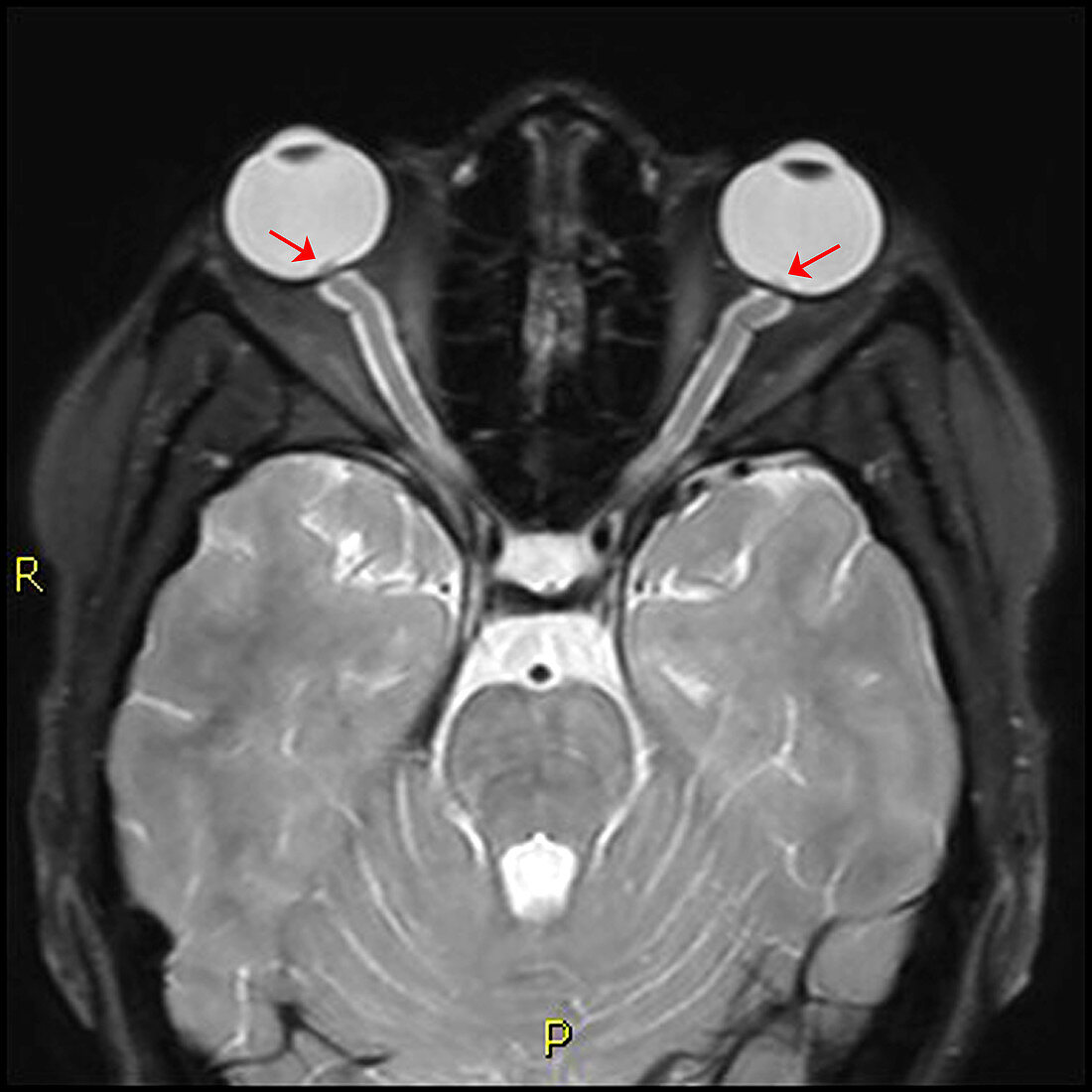
| This axial (cross sectional) T2-weighted MR image of the head at the level of the globes (eyeballs) demonstrates cupping also referred to as elevation of the optic discs (arrow on viewers left). This is a result of increased intracranial pressure in this patient with known diagnosis of Pseudotumour Cerebri (idiopathic intracranial hypertension). Also seen in increased cerebrospinal fluid in the optic sheaths,surrounding the optic nerves in both orbits and a partially empty sella turcica. Symptoms of this include headache,and variable visual symptoms including loss of vision | |
| Lizenzart: | Lizenzpflichtig |
| Credit: | Science Photo Library / Living Art Enterprises, LLC |
| Bildgröße: | 4200 px × 4200 px |
| Modell-Rechte: | nicht erforderlich |
| Eigentums-Rechte: | nicht erforderlich |
| Restrictions: |
|
Preise für dieses Bild ab 15 €
Universitäten & Organisationen
(Informationsmaterial Digital, Informationsmaterial Print, Lehrmaterial Digital etc.)
ab 15 €
Redaktionell
(Bücher, Bücher: Sach- und Fachliteratur, Digitale Medien (redaktionell) etc.)
ab 30 €
Werbung
(Anzeigen, Aussenwerbung, Digitale Medien, Fernsehwerbung, Karten, Werbemittel, Zeitschriften etc.)
ab 55 €
Handelsprodukte
(bedruckte Textilie, Kalender, Postkarte, Grußkarte, Verpackung etc.)
ab 75 €
Pauschalpreise
Rechtepakete für die unbeschränkte Bildnutzung in Print oder Online
ab 495 €
Keywords
- ct,
- t2,
- T2-gewichtet