Enhanced Large Meningioma on MRI
Bildnummer 12036399
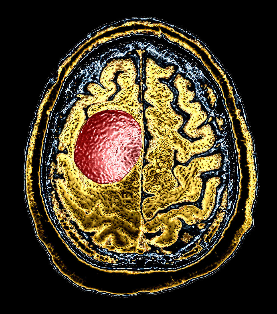
| This colour enhanced axial (cross sectional) T1-weighted MRI image of the brain shows a large,well circumscribed round mass (red) in the high posterior frontal lobe in the supplementary motor/pre-motor region compatible with a meningioma on viewers left. These account for about 20% of all intracranial masses in adults and are generally benign lesions however a few may be aggressive,infiltrating adjacent brain and growing rapidly. Occasionally they are multiple in a single patient | |
| Lizenzart: | Lizenzpflichtig |
| Credit: | Science Photo Library / Living Art Enterprises, LLC |
| Bildgröße: | 4200 px × 4757 px |
| Modell-Rechte: | nicht erforderlich |
| Eigentums-Rechte: | nicht erforderlich |
| Restrictions: |
|
Preise für dieses Bild ab 15 €
Universitäten & Organisationen
(Informationsmaterial Digital, Informationsmaterial Print, Lehrmaterial Digital etc.)
ab 15 €
Redaktionell
(Bücher, Bücher: Sach- und Fachliteratur, Digitale Medien (redaktionell) etc.)
ab 30 €
Werbung
(Anzeigen, Aussenwerbung, Digitale Medien, Fernsehwerbung, Karten, Werbemittel, Zeitschriften etc.)
ab 55 €
Handelsprodukte
(bedruckte Textilie, Kalender, Postkarte, Grußkarte, Verpackung etc.)
ab 75 €
Pauschalpreise
Rechtepakete für die unbeschränkte Bildnutzung in Print oder Online
ab 495 €