Extensive Traumatic Brain Injury CT
Bildnummer 12036390
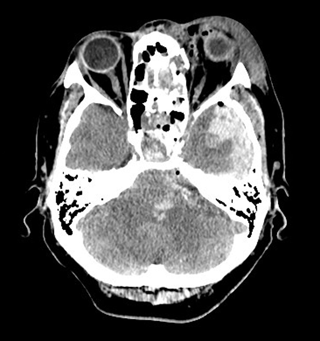
| This axial (cross sectional) CT image through the level of the orbits (eyes) shows severe soft tissue swelling/hematoma formation surrounding the eye on the viewers right. The sphenoid sinus is opacified with blood (white density) due to skull base fractures in the central part of the image. There is a subdural hematoma adjacent to the temporal lobe on viewers right in addition to temporal lobe contusions (hemorrhage-look white). Duret haemorrhages in the brainstem are seen | |
| Lizenzart: | Lizenzpflichtig |
| Credit: | Science Photo Library / Living Art Enterprises, LLC |
| Bildgröße: | 4200 px × 4475 px |
| Modell-Rechte: | nicht erforderlich |
| Eigentums-Rechte: | nicht erforderlich |
| Restrictions: |
|
Preise für dieses Bild ab 15 €
Universitäten & Organisationen
(Informationsmaterial Digital, Informationsmaterial Print, Lehrmaterial Digital etc.)
ab 15 €
Redaktionell
(Bücher, Bücher: Sach- und Fachliteratur, Digitale Medien (redaktionell) etc.)
ab 30 €
Werbung
(Anzeigen, Aussenwerbung, Digitale Medien, Fernsehwerbung, Karten, Werbemittel, Zeitschriften etc.)
ab 55 €
Handelsprodukte
(bedruckte Textilie, Kalender, Postkarte, Grußkarte, Verpackung etc.)
ab 75 €
Pauschalpreise
Rechtepakete für die unbeschränkte Bildnutzung in Print oder Online
ab 495 €