Cholesteatoma of Temporal Bone
Bildnummer 12036372
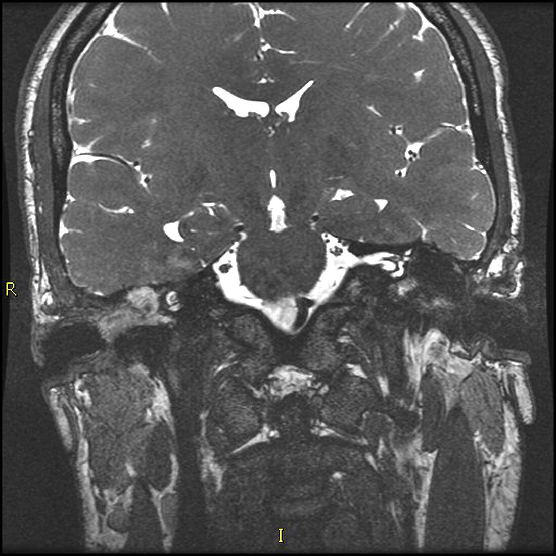
| This coronal (from the front) T2-weighted MRI image shows a destructive soft tissue lesion in the middle ear (tympanic cavity) and mastoid antrum on the viewers left as seen as an area of brighter (whiter) signal. Cholesteatomas are usually the result of a perforation (tear) of the tympanic membrane (ear drum) with ingrowth of squamous epithelial cells which shed desquamated cellular debris and keratin. However they can be congenital .These are benign lesions which progressively enlarge and are locally destructive | |
| Lizenzart: | Lizenzpflichtig |
| Credit: | Science Photo Library / Living Art Enterprises, LLC |
| Bildgröße: | 4200 px × 4200 px |
| Modell-Rechte: | nicht erforderlich |
| Eigentums-Rechte: | nicht erforderlich |
| Restrictions: |
|
Preise für dieses Bild ab 15 €
Universitäten & Organisationen
(Informationsmaterial Digital, Informationsmaterial Print, Lehrmaterial Digital etc.)
ab 15 €
Redaktionell
(Bücher, Bücher: Sach- und Fachliteratur, Digitale Medien (redaktionell) etc.)
ab 30 €
Werbung
(Anzeigen, Aussenwerbung, Digitale Medien, Fernsehwerbung, Karten, Werbemittel, Zeitschriften etc.)
ab 55 €
Handelsprodukte
(bedruckte Textilie, Kalender, Postkarte, Grußkarte, Verpackung etc.)
ab 75 €
Pauschalpreise
Rechtepakete für die unbeschränkte Bildnutzung in Print oder Online
ab 495 €