Enhanced Temporal Lobe AVM CTA
Bildnummer 12036312
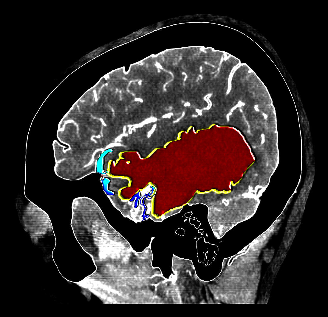
| This colour enhanced sagital (from the side) image from an intracranial CT angiogram shows a very large acute hemorrhage (bleeding) in the temporal lobe seen as the large ovoid region of red. A small collection of abnormal enhancing vessels (linear blue and turquoise densities) along the left side of the temporal lobe hemorrhage represent a high flow arterial venous malformation (AVM) which was the cause of the hemorrhge. AVMs are dangerous because of there potential for hemorrhage (bleeding) but they can also result in strokes,seizures,headaches and even death | |
| Lizenzart: | Lizenzpflichtig |
| Credit: | Science Photo Library / Living Art Enterprises, LLC |
| Bildgröße: | 4337 px × 4200 px |
| Modell-Rechte: | nicht erforderlich |
| Eigentums-Rechte: | nicht erforderlich |
| Restrictions: |
|
Preise für dieses Bild ab 15 €
Universitäten & Organisationen
(Informationsmaterial Digital, Informationsmaterial Print, Lehrmaterial Digital etc.)
ab 15 €
Redaktionell
(Bücher, Bücher: Sach- und Fachliteratur, Digitale Medien (redaktionell) etc.)
ab 30 €
Werbung
(Anzeigen, Aussenwerbung, Digitale Medien, Fernsehwerbung, Karten, Werbemittel, Zeitschriften etc.)
ab 55 €
Handelsprodukte
(bedruckte Textilie, Kalender, Postkarte, Grußkarte, Verpackung etc.)
ab 75 €
Pauschalpreise
Rechtepakete für die unbeschränkte Bildnutzung in Print oder Online
ab 495 €