Haemorrhagic Arachnoid Cyst
Bildnummer 12036282
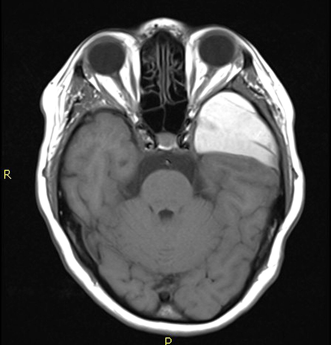
| This axial (cross sectional) T1-weighted MRI image of the brain without contrast demonstrates the typical appearance of an arachnoid cyst in the middle cranial fossa (on the viewers right) however this person sustained a head injury resulting in hemorrhage/bleeding into this extra-axial cyst. Deformity/dysmorphic anterior left (on viewers right) temporal lobe adjacent to the arachnoid cyst would be expected whether there had or had not been bleeding into the cyst | |
| Lizenzart: | Lizenzpflichtig |
| Credit: | Science Photo Library / Living Art Enterprises, LLC |
| Bildgröße: | 4200 px × 4378 px |
| Modell-Rechte: | nicht erforderlich |
| Eigentums-Rechte: | nicht erforderlich |
| Restrictions: |
|
Preise für dieses Bild ab 15 €
Universitäten & Organisationen
(Informationsmaterial Digital, Informationsmaterial Print, Lehrmaterial Digital etc.)
ab 15 €
Redaktionell
(Bücher, Bücher: Sach- und Fachliteratur, Digitale Medien (redaktionell) etc.)
ab 30 €
Werbung
(Anzeigen, Aussenwerbung, Digitale Medien, Fernsehwerbung, Karten, Werbemittel, Zeitschriften etc.)
ab 55 €
Handelsprodukte
(bedruckte Textilie, Kalender, Postkarte, Grußkarte, Verpackung etc.)
ab 75 €
Pauschalpreise
Rechtepakete für die unbeschränkte Bildnutzung in Print oder Online
ab 495 €