Cervical Myelogram (X-ray)
Bildnummer 12035627
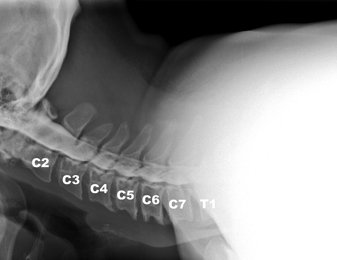
| This lateral (from the side) x-ray view of the neck demonstrates what a cervical myelogram looks like. This procedure is being done less frequently because of the widespread utilization of MRI,however they still are being used for instances when patients with pacemakers can't have an MRI or when the findings on the MRI are inconclusive. This test is obtained by first doing a spinal tap in the lower back (lumbar) region. Then a contrast agent (dye) is injected where the spinal fluid is located. When the contrast is seen in the neck,X-rays are obtained. This view shows compression along the ventral (anterior) front of the thecal sac which contains the dye secondary to multilevel disc herniations and associated bony spurring | |
| Lizenzart: | Lizenzpflichtig |
| Credit: | Science Photo Library / Living Art Enterprises |
| Bildgröße: | 5453 px × 4200 px |
| Modell-Rechte: | nicht erforderlich |
| Eigentums-Rechte: | nicht erforderlich |
| Restrictions: |
|
Preise für dieses Bild ab 15 €
Universitäten & Organisationen
(Informationsmaterial Digital, Informationsmaterial Print, Lehrmaterial Digital etc.)
ab 15 €
Redaktionell
(Bücher, Bücher: Sach- und Fachliteratur, Digitale Medien (redaktionell) etc.)
ab 30 €
Werbung
(Anzeigen, Aussenwerbung, Digitale Medien, Fernsehwerbung, Karten, Werbemittel, Zeitschriften etc.)
ab 55 €
Handelsprodukte
(bedruckte Textilie, Kalender, Postkarte, Grußkarte, Verpackung etc.)
ab 75 €
Pauschalpreise
Rechtepakete für die unbeschränkte Bildnutzung in Print oder Online
ab 495 €
Keywords
- abnormal,
- Anomalie,
- Bildgebung,
- Degeneration,
- degenerativ,
- Diagnose,
- Gebärmutterhals-,
- Hals,
- Knochen,
- Kompression,
- medizinisch,
- Myelogramm,
- Myelographie,
- Nackenschmerzen,
- Radiographie,
- Radiologie,
- Röntgen,
- Schmerz,
- Seitenansicht,
- seitlich,
- Skelett-,
- ungesund,
- Wirbel,
- Wirbelsäule,
- Wirbelsäulen-,
- Wissenschaft