Invasive Squamous Carcinoma (LM)
Bildnummer 12028893
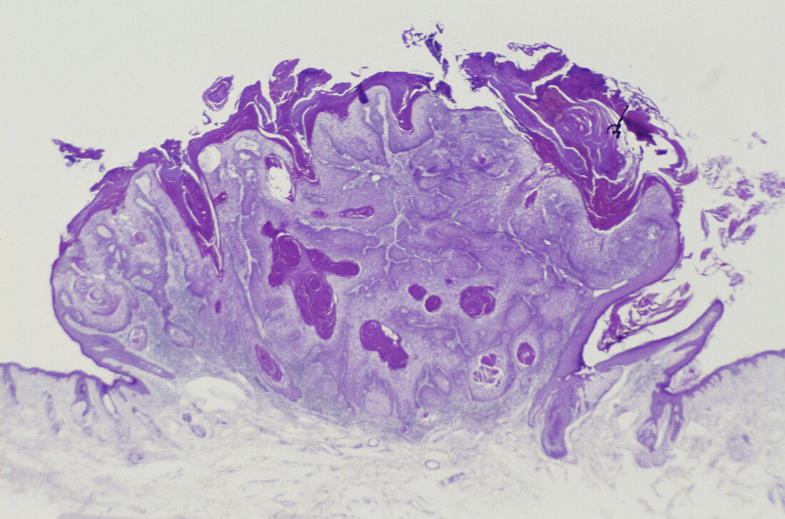
| Light micrograph (LM) showing invasive squamous carcinoma. In a well-differentiated squamous carcinoma,as shown in this example from the pinna,there may be hyperkeratosis,parakeratotis,or crusting ulceration over an area of proliferating keratinocytes. Cytologically,the cells exhibit features of atypica with plenomorphism,nuclear hyperchromatism,and increase in mitoses. The main feature of this lesion is invasion of the dermis by the proliferating cells. Magnification unknown | |
| Lizenzart: | Lizenzpflichtig |
| Credit: | Science Photo Library / Biophoto Associates |
| Bildgröße: | 5141 px × 3399 px |
| Modell-Rechte: | nicht erforderlich |
| Eigentums-Rechte: | nicht erforderlich |
| Restrictions: |
|
Preise für dieses Bild ab 15 €
Universitäten & Organisationen
(Informationsmaterial Digital, Informationsmaterial Print, Lehrmaterial Digital etc.)
ab 15 €
Redaktionell
(Bücher, Bücher: Sach- und Fachliteratur, Digitale Medien (redaktionell) etc.)
ab 30 €
Werbung
(Anzeigen, Aussenwerbung, Digitale Medien, Fernsehwerbung, Karten, Werbemittel, Zeitschriften etc.)
ab 55 €
Handelsprodukte
(bedruckte Textilie, Kalender, Postkarte, Grußkarte, Verpackung etc.)
ab 75 €
Pauschalpreise
Rechtepakete für die unbeschränkte Bildnutzung in Print oder Online
ab 495 €