Illustration of Spinal Disk Pathologies
Bildnummer 12026302
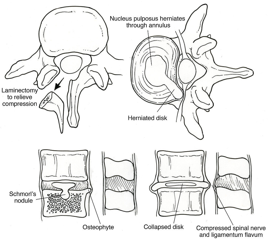
| Anatomical illustration of spinal disk pathologies,showing (upper left) a laminectomy to relieve cord compression,(upper right) herniated disk,where the nucleus pulposus herniates through annulus,and (bottom row,left),a Schmorl's nodule protruding into osteophyte and (bottom row,right) a collapsed disk with compressed spinal nerve and ligamentum flavum | |
| Lizenzart: | Lizenzpflichtig |
| Credit: | Science Photo Library / Science Source |
| Bildgröße: | 3654 px × 3412 px |
| Modell-Rechte: | nicht erforderlich |
| Eigentums-Rechte: | nicht erforderlich |
| Restrictions: |
|
Preise für dieses Bild ab 15 €
Universitäten & Organisationen
(Informationsmaterial Digital, Informationsmaterial Print, Lehrmaterial Digital etc.)
ab 15 €
Redaktionell
(Bücher, Bücher: Sach- und Fachliteratur, Digitale Medien (redaktionell) etc.)
ab 30 €
Werbung
(Anzeigen, Aussenwerbung, Digitale Medien, Fernsehwerbung, Karten, Werbemittel, Zeitschriften etc.)
ab 55 €
Handelsprodukte
(bedruckte Textilie, Kalender, Postkarte, Grußkarte, Verpackung etc.)
ab 75 €
Pauschalpreise
Rechtepakete für die unbeschränkte Bildnutzung in Print oder Online
ab 495 €