Large Infantile Stroke,MRI
Bildnummer 12013639
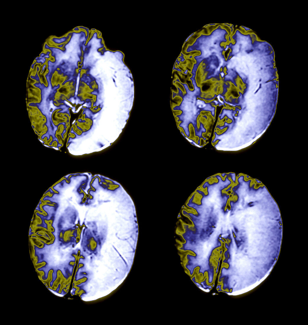
| This enhanced composite of 4 axial,(cross sectional),T2 weighted MRI images of the brain in an infant shows a very large infarct,(stroke),in the middle cerebral artery vascular territory. This is the large wedge-shaped region of abnormal increased signal,(looks white),on the left side of the brain,(your right). The right side of the brain,which is unaffected,shows differentiation of cortical gray matter,(darker band around the brain),from the immature,unmyelinated white matter which looks white on this sequence due to the large amount of water in unmyelinated regions of the white matter | |
| Lizenzart: | Lizenzpflichtig |
| Credit: | Science Photo Library / Living Art Enterprises |
| Bildgröße: | 3600 px × 3795 px |
| Modell-Rechte: | nicht erforderlich |
| Eigentums-Rechte: | nicht erforderlich |
| Restrictions: |
|
Preise für dieses Bild ab 15 €
Universitäten & Organisationen
(Informationsmaterial Digital, Informationsmaterial Print, Lehrmaterial Digital etc.)
ab 15 €
Redaktionell
(Bücher, Bücher: Sach- und Fachliteratur, Digitale Medien (redaktionell) etc.)
ab 30 €
Werbung
(Anzeigen, Aussenwerbung, Digitale Medien, Fernsehwerbung, Karten, Werbemittel, Zeitschriften etc.)
ab 55 €
Handelsprodukte
(bedruckte Textilie, Kalender, Postkarte, Grußkarte, Verpackung etc.)
ab 75 €
Pauschalpreise
Rechtepakete für die unbeschränkte Bildnutzung in Print oder Online
ab 495 €