MR of Malignant Brain Tumor,3 of 3
Bildnummer 12010570
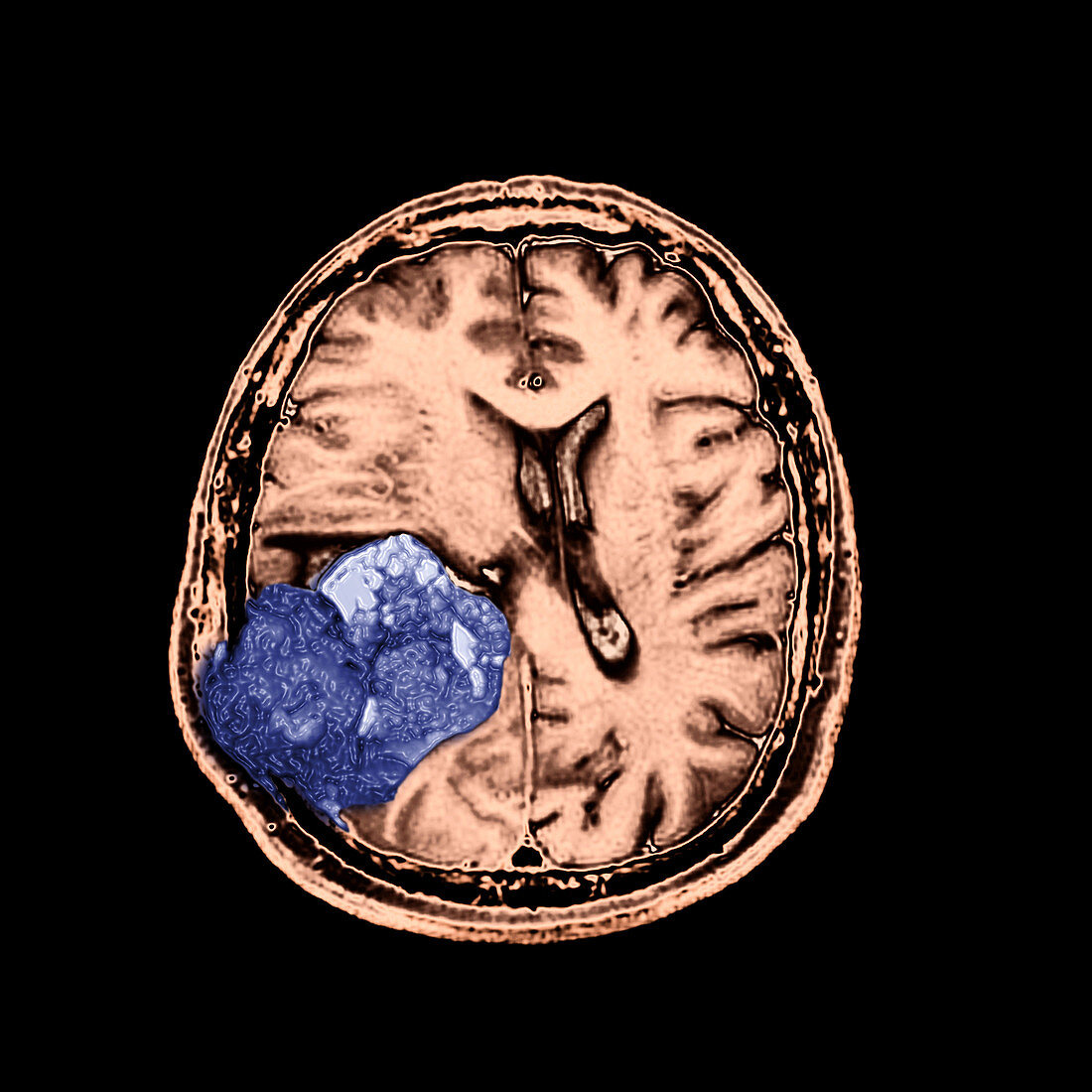
| This color enhanced axial (cross sectional) T2 weighted image of the brain demonstrates a heterogenous tumor (blue) in the right (your left) parietal lobe which destroys the overlying calvarium (skull bone) and extends into the overlying scalp. This tumor represents a malignant form of an astrocytoma. It is a WHO grade IV tumor called a Glioblasstoma Multiforme (GBM). These are most commonly seen in the elderly and unfortunately are one of the most common astrocytomas. They carry a poor,short term survival. Image 3 of 3 | |
| Lizenzart: | Lizenzpflichtig |
| Credit: | Science Photo Library / Living Art Enterprises |
| Bildgröße: | 3600 px × 3600 px |
| Modell-Rechte: | nicht erforderlich |
| Eigentums-Rechte: | nicht erforderlich |
| Restrictions: |
|
Preise für dieses Bild ab 15 €
Universitäten & Organisationen
(Informationsmaterial Digital, Informationsmaterial Print, Lehrmaterial Digital etc.)
ab 15 €
Redaktionell
(Bücher, Bücher: Sach- und Fachliteratur, Digitale Medien (redaktionell) etc.)
ab 30 €
Werbung
(Anzeigen, Aussenwerbung, Digitale Medien, Fernsehwerbung, Karten, Werbemittel, Zeitschriften etc.)
ab 55 €
Handelsprodukte
(bedruckte Textilie, Kalender, Postkarte, Grußkarte, Verpackung etc.)
ab 75 €
Pauschalpreise
Rechtepakete für die unbeschränkte Bildnutzung in Print oder Online
ab 495 €