Cerebral Angioma
Bildnummer 12010499
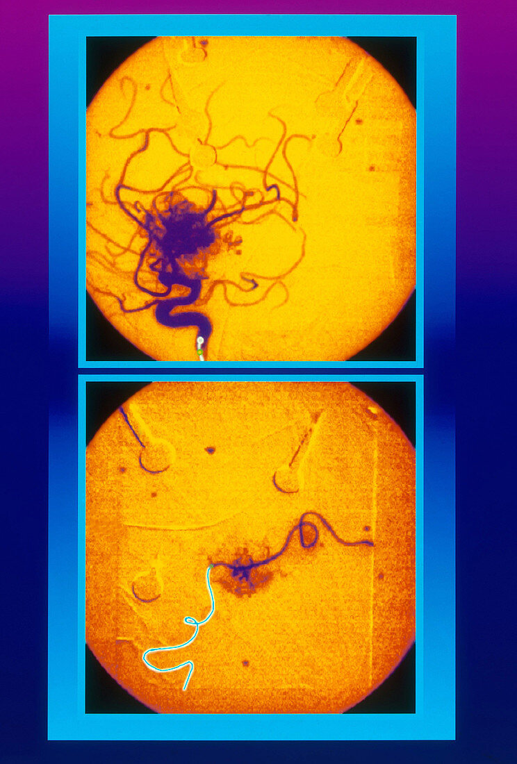
| Angioma case using FasTRACKER-18 system. Magenta to blue background with intermediary panel in cyan and blue. Two rectangular procedural panels,one above the other,with anatomy vignetted in yellow-orange,each panel with cyan borders. Top panel visualizes angioma in orange and purple-blue. Lower panel visualizes introduction of catheter in cyan with blue guide-wire to site of origin of angioma,showing successful reduction of perfused vessels | |
| Lizenzart: | Lizenzpflichtig |
| Credit: | Science Photo Library / Lunagrafix |
| Bildgröße: | 2558 px × 3784 px |
| Modell-Rechte: | nicht erforderlich |
| Eigentums-Rechte: | nicht erforderlich |
| Restrictions: |
|
Preise für dieses Bild ab 15 €
Universitäten & Organisationen
(Informationsmaterial Digital, Informationsmaterial Print, Lehrmaterial Digital etc.)
ab 15 €
Redaktionell
(Bücher, Bücher: Sach- und Fachliteratur, Digitale Medien (redaktionell) etc.)
ab 30 €
Werbung
(Anzeigen, Aussenwerbung, Digitale Medien, Fernsehwerbung, Karten, Werbemittel, Zeitschriften etc.)
ab 55 €
Handelsprodukte
(bedruckte Textilie, Kalender, Postkarte, Grußkarte, Verpackung etc.)
ab 75 €
Pauschalpreise
Rechtepakete für die unbeschränkte Bildnutzung in Print oder Online
ab 495 €