MRI of Acute MS
Bildnummer 12006787
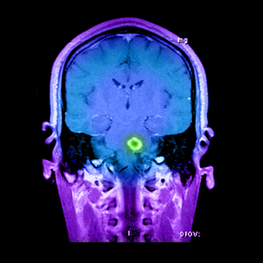
| This coronal (frontal) contrast enhanced MRI image of the brain through the level of the brainstem reveals a prominent ring enhancing lesion in the left side of the pons (green). T2 weighted images demonstrated extensive,mass-like brainstem edema and swelling. This represents a form of acute MS that can stimulate a tumor therefore it is referred to as tumefactive | |
| Lizenzart: | Lizenzpflichtig |
| Credit: | Science Photo Library / Medical Body Scans |
| Bildgröße: | 3600 px × 3600 px |
| Modell-Rechte: | nicht erforderlich |
| Eigentums-Rechte: | nicht erforderlich |
| Restrictions: |
|
Preise für dieses Bild ab 15 €
Universitäten & Organisationen
(Informationsmaterial Digital, Informationsmaterial Print, Lehrmaterial Digital etc.)
ab 15 €
Redaktionell
(Bücher, Bücher: Sach- und Fachliteratur, Digitale Medien (redaktionell) etc.)
ab 30 €
Werbung
(Anzeigen, Aussenwerbung, Digitale Medien, Fernsehwerbung, Karten, Werbemittel, Zeitschriften etc.)
ab 55 €
Handelsprodukte
(bedruckte Textilie, Kalender, Postkarte, Grußkarte, Verpackung etc.)
ab 75 €
Pauschalpreise
Rechtepakete für die unbeschränkte Bildnutzung in Print oder Online
ab 495 €