Oligodendroglioma
Bildnummer 12006772
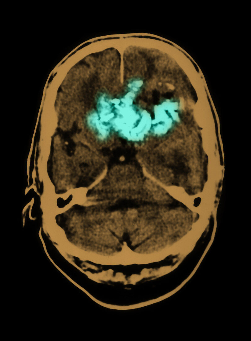
| This axial (cross sectional) CT image of the brain show a large,prominently calcified,heterogenous frontal lobe mass (shown in light green) with associated mass effect. There is surrounding edema.This is a primary brain tumor called an oligodendroglioma.This is a type of primary intracranial tumor called a glioma which arise from the supporting elements. In this case the oligodendrocytes are glial cells which normally are involved with the formation of myelin. These types of tumors have a very high incidence of calcifications. This is a colorized version of BF3177 | |
| Lizenzart: | Lizenzpflichtig |
| Credit: | Science Photo Library / Medical Body Scans |
| Bildgröße: | 5395 px × 7324 px |
| Modell-Rechte: | nicht erforderlich |
| Eigentums-Rechte: | nicht erforderlich |
| Restrictions: |
|
Preise für dieses Bild ab 15 €
Universitäten & Organisationen
(Informationsmaterial Digital, Informationsmaterial Print, Lehrmaterial Digital etc.)
ab 15 €
Redaktionell
(Bücher, Bücher: Sach- und Fachliteratur, Digitale Medien (redaktionell) etc.)
ab 30 €
Werbung
(Anzeigen, Aussenwerbung, Digitale Medien, Fernsehwerbung, Karten, Werbemittel, Zeitschriften etc.)
ab 55 €
Handelsprodukte
(bedruckte Textilie, Kalender, Postkarte, Grußkarte, Verpackung etc.)
ab 75 €
Pauschalpreise
Rechtepakete für die unbeschränkte Bildnutzung in Print oder Online
ab 495 €