Embryo at 26 days
Bildnummer 11999829
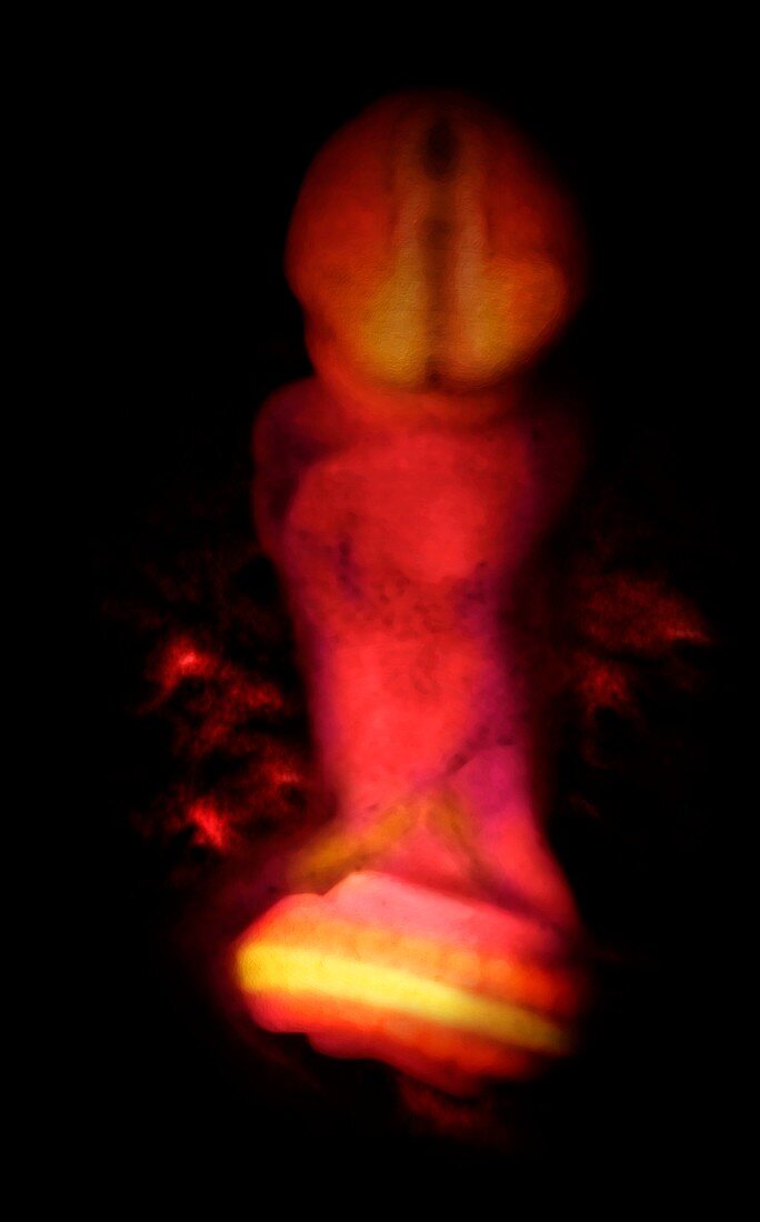
| Embryo at 26 days,computer-generated image from a micro-MRI scan. At this stage the embryo measures 4 millimetres in length. The head region (top) is bent downwards so that the developing central nervous system can be seen. The tube-like structure in the head region indicates the developing spinal cord. The tail of the embryo can be seen curving upwards so that the spinal cord and somites (muscle blocks,orange) are seen | |
| Lizenzart: | Lizenzpflichtig |
| Credit: | Science Photo Library / Anatomical Travelogue |
| Bildgröße: | 3319 px × 5334 px |
| Modell-Rechte: | nicht erforderlich |
| Eigentums-Rechte: | nicht erforderlich |
| Restrictions: |
|
Preise für dieses Bild ab 15 €
Universitäten & Organisationen
(Informationsmaterial Digital, Informationsmaterial Print, Lehrmaterial Digital etc.)
ab 15 €
Redaktionell
(Bücher, Bücher: Sach- und Fachliteratur, Digitale Medien (redaktionell) etc.)
ab 30 €
Werbung
(Anzeigen, Aussenwerbung, Digitale Medien, Fernsehwerbung, Karten, Werbemittel, Zeitschriften etc.)
ab 55 €
Handelsprodukte
(bedruckte Textilie, Kalender, Postkarte, Grußkarte, Verpackung etc.)
ab 75 €
Pauschalpreise
Rechtepakete für die unbeschränkte Bildnutzung in Print oder Online
ab 495 €