Breathing tube in fruit fly puparium
Bildnummer 11908386
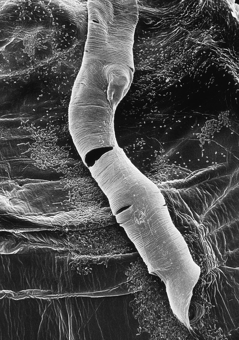
| Scanning electron micrograph of the inner surface of a pupal case,or puparium,of the fruit fly Drosophila melanogaster (wild type Oregon R). Seen is a split tracheal tube,forming part of the respiratory apparatus of the pupa. Tracheal tubes carry air,entering through the spiracles,(breathing tubes) around the puparium. They are made of chitin & constructed in a spiral pattern. The respiratory apparatus (spiracles & tracheae) is not integral to the pupa,but forms part of the puparium. It is shed with the case at eclosion (hatching). Numerous yeast cells are seen on the surface of the puparium. Magnificaton: x700 at 10x8 inch size. size,x90 at 35mm size | |
| Lizenzart: | Lizenzpflichtig |
| Credit: | Science Photo Library / Burgess, Dr. Jeremy |
| Bildgröße: | 3543 px × 5045 px |
| Modell-Rechte: | nicht erforderlich |
| Eigentums-Rechte: | nicht erforderlich |
| Restrictions: | - |
Preise für dieses Bild ab 15 €
Universitäten & Organisationen
(Informationsmaterial Digital, Informationsmaterial Print, Lehrmaterial Digital etc.)
ab 15 €
Redaktionell
(Bücher, Bücher: Sach- und Fachliteratur, Digitale Medien (redaktionell) etc.)
ab 30 €
Werbung
(Anzeigen, Aussenwerbung, Digitale Medien, Fernsehwerbung, Karten, Werbemittel, Zeitschriften etc.)
ab 55 €
Handelsprodukte
(bedruckte Textilie, Kalender, Postkarte, Grußkarte, Verpackung etc.)
ab 75 €
Pauschalpreise
Rechtepakete für die unbeschränkte Bildnutzung in Print oder Online
ab 495 €