Intestinal protozoan parasites,TEM
Bildnummer 11905957
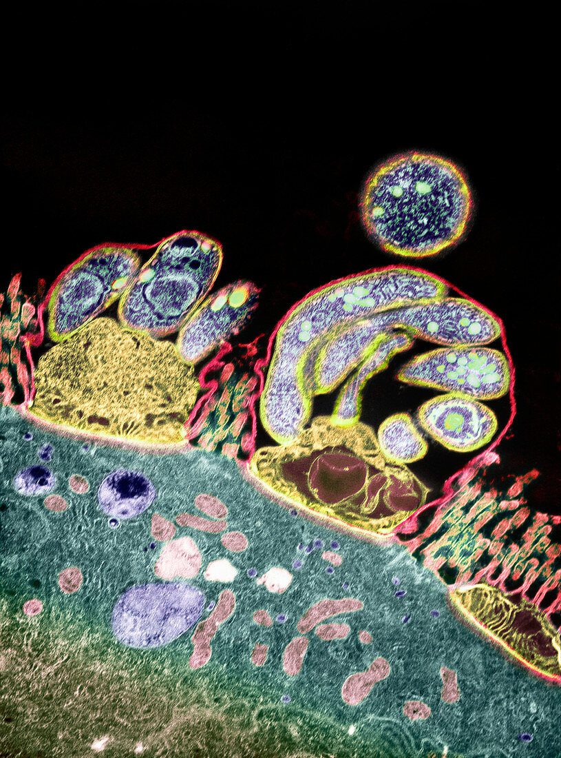
| Parasitic protozoa on intestinal surface. Coloured transmission electron micrograph (TEM) of several meront stages of Cryptosporidium sp. protozoan parasites (round) on an intestinal surface. The surface is covered in microvilli strands. Meronts feed through the yellow structures attaching them to the surface. The meront stage of the life cycle involves asexual reproduction to form merozoites (blue). When released,the merozoites may repeat asexual reproduction,or may reproduce sexually to form the oocyst stage that passes between hosts in infected faeces. This parasite causes diarrhoea. Magnification: x8300 when printed 10cm high | |
| Lizenzart: | Lizenzpflichtig |
| Credit: | Science Photo Library / London School of Hygiene & Tropical Medicine |
| Bildgröße: | 2500 px × 3375 px |
| Modell-Rechte: | nicht erforderlich |
| Eigentums-Rechte: | nicht erforderlich |
| Restrictions: | - |
Preise für dieses Bild ab 15 €
Universitäten & Organisationen
(Informationsmaterial Digital, Informationsmaterial Print, Lehrmaterial Digital etc.)
ab 15 €
Redaktionell
(Bücher, Bücher: Sach- und Fachliteratur, Digitale Medien (redaktionell) etc.)
ab 30 €
Werbung
(Anzeigen, Aussenwerbung, Digitale Medien, Fernsehwerbung, Karten, Werbemittel, Zeitschriften etc.)
ab 55 €
Handelsprodukte
(bedruckte Textilie, Kalender, Postkarte, Grußkarte, Verpackung etc.)
ab 75 €
Pauschalpreise
Rechtepakete für die unbeschränkte Bildnutzung in Print oder Online
ab 495 €
Keywords
- Aids,
- asexuell,
- Bühne,
- Cryptosporidium sp.,
- Darm,
- Darm-,
- Durchfall,
- elektronenmikroskopische Aufnahme,
- Erreger,
- farbig,
- Fütterung,
- Gedärme,
- Gesundheitswesen,
- HIV,
- Infektion,
- infiziert,
- Kondition,
- kopierend,
- Krankheit,
- Krankheitserreger,
- Lebenszyklus,
- Medizin,
- medizinisch,
- merozoiten,
- Mikrobe,
- Mikroben,
- Mikrobiologie,
- mikrobiologisch,
- Mikroorganismen,
- Mikroorganismus,
- Mikrovilli,
- Natur,
- Parasit,
- parasitär,
- Parasiten,
- pathogen,
- Protozoen,
- Protozoon,
- Replikation,
- Reproduktion,
- reproduktiv,
- Schizogonie,
- Sektion,
- sektioniert,
- Störung,
- Struktur,
- tem,
- Tier,
- Tierwelt,
- Übertragung,
- Verdauung,
- Verdauungs-,
- wirbellos,
- Wirbellose,
- Zoologie