Abdomen,CT scan
Bildnummer 11877114
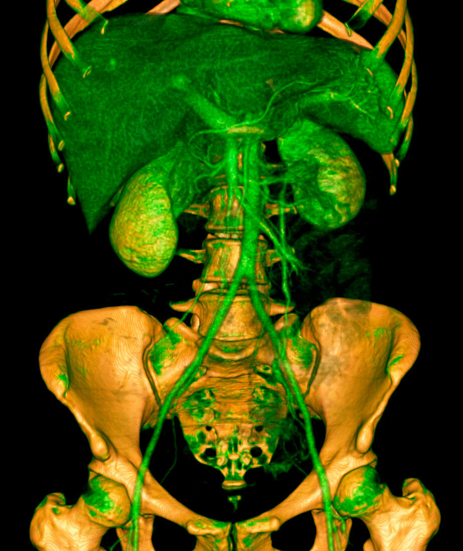
| Abdomen,CT scan. Coloured three-dimensional computed tomography (CT) scan of an abdomen in frontal view. The image shows (top to bottom),rib cage,liver,kidneys,lower spine,abdominal aorta,pelvis and femur joints. This image was produced using a multi-slice CT scanner,which uses a thin X-ray beam to scan around the patient to create 'slices' of the body. A computer reconstructs the slices into coloured three- dimensional images including bones and soft tissue. A contrast agent,injected into the patient,enables blood vessels to be detected by the CT scanner. The surgeon can navigate through the data by using a 'fly-through' animation of the images. This image was created using OsiriX medical imaging software | |
| Lizenzart: | Lizenzpflichtig |
| Credit: | Science Photo Library / Rosset, Antoine |
| Bildgröße: | 2719 px × 3236 px |
| Modell-Rechte: | nicht erforderlich |
| Eigentums-Rechte: | nicht erforderlich |
| Restrictions: | - |
Preise für dieses Bild ab 15 €
Universitäten & Organisationen
(Informationsmaterial Digital, Informationsmaterial Print, Lehrmaterial Digital etc.)
ab 15 €
Redaktionell
(Bücher, Bücher: Sach- und Fachliteratur, Digitale Medien (redaktionell) etc.)
ab 30 €
Werbung
(Anzeigen, Aussenwerbung, Digitale Medien, Fernsehwerbung, Karten, Werbemittel, Zeitschriften etc.)
ab 55 €
Handelsprodukte
(bedruckte Textilie, Kalender, Postkarte, Grußkarte, Verpackung etc.)
ab 75 €
Pauschalpreise
Rechtepakete für die unbeschränkte Bildnutzung in Print oder Online
ab 495 €
Keywords
- 3-d,
- 3D,
- Anatomie,
- anatomisch,
- Angiogramm,
- ärztliche Untersuchung,
- Computertomographie,
- CT-Scan,
- CTA,
- Diagnose,
- Dreidimensional,
- farbig,
- gefärbt,
- innere Organe,
- Knochen,
- Kontrastmittel,
- Medizin,
- medizinisch,
- medizinische Bildgebung,
- medizinische Visualisierung,
- medizinischer Scan,
- Mensch,
- Menschen,
- menschlicher Körper,
- Organ,
- Organe,
- OsiriX,
- Person,
- Querschnitt,
- Radiographie,
- Radiologie,
- radiologisch,
- Röntgen,
- Röntgenstrahlen,
- Röntgenstrahlung,
- Scanner