Thorax and abdomen,EBT scan
Bildnummer 11877110
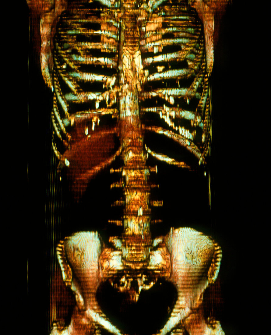
| Thorax and abdomen. Coloured 3-D electron beam tomography (EBT) scan of a male thoracic cavity & abdomen. EBT provides clearer and more detailed information about body tissues than CT (computed tomography) scans & reduces the time patients are exposed to dangerous radiation. EBT involves firing an electron beam against tungsten targets arranged in a 210 degree arc below the patient. These produce X-rays that pass through the patient. A computer produces cross-sectional (slice) images of the body tissues,and 3-D images can be built up by combining many slices. The ribs (upper centre),vertebral column (down centre) and pelvis (hips,lower centre) are seen | |
| Lizenzart: | Lizenzpflichtig |
| Credit: | Science Photo Library / King-Holmes, James |
| Bildgröße: | 2873 px × 3551 px |
| Modell-Rechte: | nicht erforderlich |
| Eigentums-Rechte: | nicht erforderlich |
| Restrictions: | - |
Preise für dieses Bild ab 15 €
Universitäten & Organisationen
(Informationsmaterial Digital, Informationsmaterial Print, Lehrmaterial Digital etc.)
ab 15 €
Redaktionell
(Bücher, Bücher: Sach- und Fachliteratur, Digitale Medien (redaktionell) etc.)
ab 30 €
Werbung
(Anzeigen, Aussenwerbung, Digitale Medien, Fernsehwerbung, Karten, Werbemittel, Zeitschriften etc.)
ab 55 €
Handelsprodukte
(bedruckte Textilie, Kalender, Postkarte, Grußkarte, Verpackung etc.)
ab 75 €
Pauschalpreise
Rechtepakete für die unbeschränkte Bildnutzung in Print oder Online
ab 495 €
Keywords
- 3-d,
- Abdomen,
- Anatomie,
- Bauch,
- Becken,
- Bild,
- Bildgebung,
- Diagnose,
- Dreidimensional,
- EBCT,
- ebt,
- England,
- farbig,
- Gesundheitswesen,
- Hohlraum,
- Hüfte,
- Hüften,
- Käfig,
- London,
- Medizin,
- medizinisch,
- menschlicher Körper,
- Radiographie,
- Rippe,
- Rippen,
- Röntgen,
- Scan,
- Steißbein,
- Sternum,
- Technik,
- Technologie,
- technologisch,
- thorakal,
- Thorax,
- Torso,
- Truhe,
- Wirbel,
- Wirbelsäule