MRI child body scan
Bildnummer 11877094
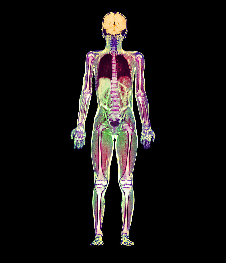
| Whole body scan. Coloured magnetic resonance imaging (MRI) whole body scan of a nine year old boy in coronal (frontal) section. His brain (orange) is seen at upper centre. The skeleton (white/purple) is visible as the long bones of the limbs and the vertebrae of the spine. In the chest,the lungs are dark purple. In the abdomen,the lobes of the liver are green & purple,while the oval bladder (dark purple) is in the pelvis. This whole body image is the product of a number of MRI scans made along the length of the body and then combined. MRI scanning uses radio waves and a powerful magnetic field to produce "slice" images through the body | |
| Lizenzart: | Lizenzpflichtig |
| Credit: | Science Photo Library / Fraser, Simon |
| Bildgröße: | 2400 px × 2800 px |
| Modell-Rechte: | nicht erforderlich |
| Eigentums-Rechte: | nicht erforderlich |
| Restrictions: | - |
Preise für dieses Bild ab 15 €
Universitäten & Organisationen
(Informationsmaterial Digital, Informationsmaterial Print, Lehrmaterial Digital etc.)
ab 15 €
Redaktionell
(Bücher, Bücher: Sach- und Fachliteratur, Digitale Medien (redaktionell) etc.)
ab 30 €
Werbung
(Anzeigen, Aussenwerbung, Digitale Medien, Fernsehwerbung, Karten, Werbemittel, Zeitschriften etc.)
ab 55 €
Handelsprodukte
(bedruckte Textilie, Kalender, Postkarte, Grußkarte, Verpackung etc.)
ab 75 €
Pauschalpreise
Rechtepakete für die unbeschränkte Bildnutzung in Print oder Online
ab 495 €