MRI body scan
Bildnummer 11877088
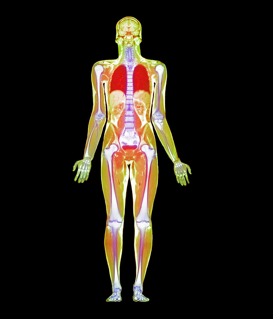
| Whole body scan. Coloured magnetic resonance imaging (MRI) scan of the whole body of a man in coronal (frontal) section. His skeleton (white) is visible as the long bones of the limbs and the vertebrae of the spine. At top,the two cerebral hemispheres of the brain are seen in the skull. In the chest,the lungs are red. In the abdomen,the lobes of the liver are light brown,while the flattened bladder (red) is in the pelvis. This whole body image is the product of a number of MRI scans made along the length of the body and then combined. MRI scanning uses radio waves and a powerful magnetic field to produce "slice" images through the body | |
| Lizenzart: | Lizenzpflichtig |
| Credit: | Science Photo Library / Fraser, Simon |
| Bildgröße: | 2400 px × 2800 px |
| Modell-Rechte: | nicht erforderlich |
| Eigentums-Rechte: | nicht erforderlich |
| Restrictions: | - |
Preise für dieses Bild ab 15 €
Universitäten & Organisationen
(Informationsmaterial Digital, Informationsmaterial Print, Lehrmaterial Digital etc.)
ab 15 €
Redaktionell
(Bücher, Bücher: Sach- und Fachliteratur, Digitale Medien (redaktionell) etc.)
ab 30 €
Werbung
(Anzeigen, Aussenwerbung, Digitale Medien, Fernsehwerbung, Karten, Werbemittel, Zeitschriften etc.)
ab 55 €
Handelsprodukte
(bedruckte Textilie, Kalender, Postkarte, Grußkarte, Verpackung etc.)
ab 75 €
Pauschalpreise
Rechtepakete für die unbeschränkte Bildnutzung in Print oder Online
ab 495 €