MRI scan of a whole human body (female)
Bildnummer 11877085
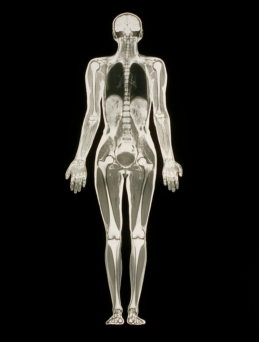
| Whole body scan. Magnetic resonance imaging (MRI) scan of the whole body of a woman,in coronal (frontal) section. Various parts of the anatomy of the human body are seen. The skeleton is visible as white long bones of the limbs and vertebrae of the spine. At top,the two cerebral hemispheres of the brain are seen. In the chest,the lungs are black. In the abdomen,lobes of the liver are grey,while the bladder appears dark and rounded in the pelvis. This whole body image is the product of a number of MRI scans made along the length of the body and combined. MRI scanning uses radio waves and a powerful magnetic field to produce "slice" images through the body | |
| Lizenzart: | Lizenzpflichtig |
| Credit: | Science Photo Library / Fraser, Simon |
| Bildgröße: | 3550 px × 4669 px |
| Modell-Rechte: | nicht erforderlich |
| Eigentums-Rechte: | nicht erforderlich |
| Restrictions: | - |
Preise für dieses Bild ab 15 €
Universitäten & Organisationen
(Informationsmaterial Digital, Informationsmaterial Print, Lehrmaterial Digital etc.)
ab 15 €
Redaktionell
(Bücher, Bücher: Sach- und Fachliteratur, Digitale Medien (redaktionell) etc.)
ab 30 €
Werbung
(Anzeigen, Aussenwerbung, Digitale Medien, Fernsehwerbung, Karten, Werbemittel, Zeitschriften etc.)
ab 55 €
Handelsprodukte
(bedruckte Textilie, Kalender, Postkarte, Grußkarte, Verpackung etc.)
ab 75 €
Pauschalpreise
Rechtepakete für die unbeschränkte Bildnutzung in Print oder Online
ab 495 €