False-col NMR section through adult female pelvis
Bildnummer 11877071
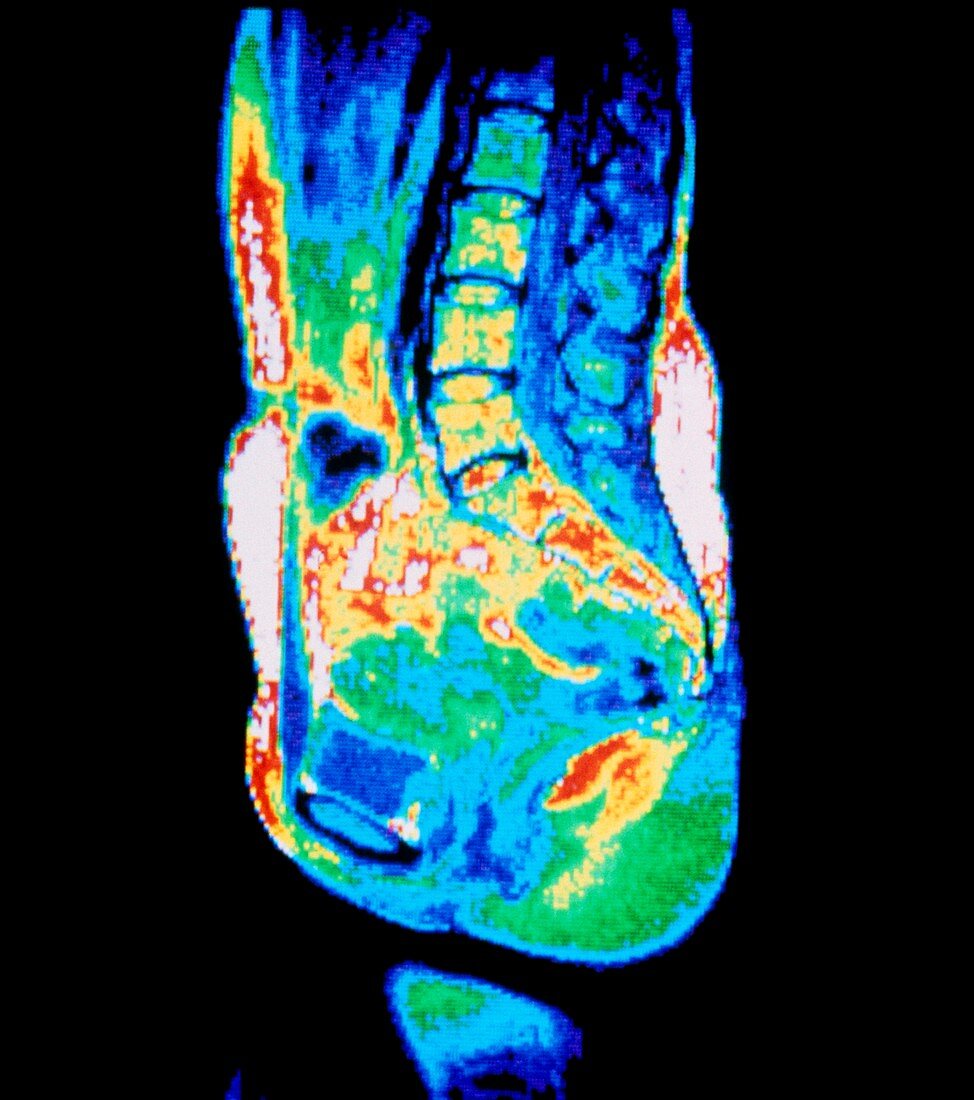
| False colour nuclear magnetic resonance image (NMR scan) of a median (mid-sagittal) section through a normal,adult female pelvis (front is at left). Vertebrae in the lumbar spine and coccyx are visible,with the intervertebral discs of the lumbar spine apparent and bones of the coccyx coded red. The green area at far bottom right corresponds to the muscles of the buttocks. Other structures seen include the pubic symphysis (the joint between the pubic bones of the pelvis),which appears as a light blue oval shape at bottom left | |
| Lizenzart: | Lizenzpflichtig |
| Credit: | Science Photo Library / CNRI |
| Bildgröße: | 3934 px × 4441 px |
| Modell-Rechte: | nicht erforderlich |
| Eigentums-Rechte: | nicht erforderlich |
| Restrictions: | - |
Preise für dieses Bild ab 15 €
Universitäten & Organisationen
(Informationsmaterial Digital, Informationsmaterial Print, Lehrmaterial Digital etc.)
ab 15 €
Redaktionell
(Bücher, Bücher: Sach- und Fachliteratur, Digitale Medien (redaktionell) etc.)
ab 30 €
Werbung
(Anzeigen, Aussenwerbung, Digitale Medien, Fernsehwerbung, Karten, Werbemittel, Zeitschriften etc.)
ab 55 €
Handelsprodukte
(bedruckte Textilie, Kalender, Postkarte, Grußkarte, Verpackung etc.)
ab 75 €
Pauschalpreise
Rechtepakete für die unbeschränkte Bildnutzung in Print oder Online
ab 495 €