Hair follicle,SEM
Bildnummer 11876533
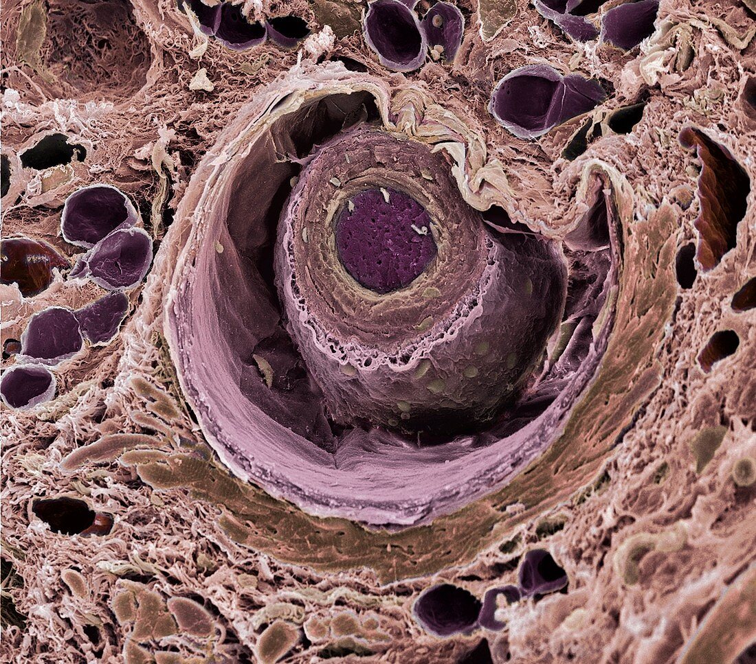
| Hair follicle. Coloured scanning electron micrograph (SEM) of a transverse freeze-fractured section through a hair shaft and follicle (round structures at centre). The internal layers of the follicle and shaft are exposed. The shaft is the innermost structure (purple) and is surrounded by its root sheath (brown/pink layers) that is part of the follicle,the structure from which a hair grows from the skin. Further outwards,there are layers of connective tissue that make up the outer part of the follicle. In the surrounding skin is the smooth muscle (dark brown arc,lower right) of the arrector pili muscle that makes a hair shaft stand erect. Magnification: x200 at 6x7cm size | |
| Lizenzart: | Lizenzpflichtig |
| Credit: | Science Photo Library / Gschmeissner, Steve |
| Bildgröße: | 4500 px × 3956 px |
| Modell-Rechte: | nicht erforderlich |
| Eigentums-Rechte: | nicht erforderlich |
| Restrictions: | - |
Preise für dieses Bild ab 15 €
Universitäten & Organisationen
(Informationsmaterial Digital, Informationsmaterial Print, Lehrmaterial Digital etc.)
ab 15 €
Redaktionell
(Bücher, Bücher: Sach- und Fachliteratur, Digitale Medien (redaktionell) etc.)
ab 30 €
Werbung
(Anzeigen, Aussenwerbung, Digitale Medien, Fernsehwerbung, Karten, Werbemittel, Zeitschriften etc.)
ab 55 €
Handelsprodukte
(bedruckte Textilie, Kalender, Postkarte, Grußkarte, Verpackung etc.)
ab 75 €
Pauschalpreise
Rechtepakete für die unbeschränkte Bildnutzung in Print oder Online
ab 495 €
Keywords
- Anatomie,
- Bilder,
- Bindegewebe,
- dermal,
- diagonal,
- Fächer,
- farbig,
- Follikel,
- Fraktur,
- frakturiert,
- gefriergebrochen,
- Geschichtet,
- gesund,
- Gewebe,
- Haar,
- Haut,
- Histologie,
- Keratin,
- menschlicher Körper,
- mikroskopische Fotos,
- normal,
- Oberfläche,
- rasterelektronenmikroskopische Aufnahme,
- REM,
- Schicht,
- Schichten,
- Sektion,
- sektioniert,
- vergrößertes Bild,
- Welle,
- Wurzel