LM of a mouse embryo at the two-cell stage
Bildnummer 11875490
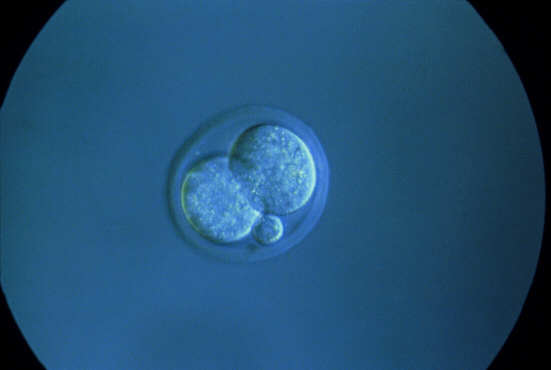
| Light micrograph of the embryo of a mouse at the two-cell stage. It is completely surrounded by a thin,resistant glycoprotein layer,the zona pellucida which a spermatozoon penetrated in order to fertilise the egg. A polar body is also here visible as a tiny sphere. At fertilisation three such cells are present together with the larger,fertilised egg cell known as the secondary oocyte. The secondary oocyte,by successive mitotic divisions,gives rise to the foetus whereas the polar bodies shortly degenerate. The embryo of a mouse reaches the four-cell stage 1.5- 2 days after ovulation and,a day after,it enters the uterus. Magnification unknown | |
| Lizenzart: | Lizenzpflichtig |
| Credit: | Science Photo Library / David Spears |
| Bildgröße: | 3692 px × 2480 px |
| Modell-Rechte: | nicht erforderlich |
| Eigentums-Rechte: | nicht erforderlich |
| Restrictions: | - |
Preise für dieses Bild ab 15 €
Universitäten & Organisationen
(Informationsmaterial Digital, Informationsmaterial Print, Lehrmaterial Digital etc.)
ab 15 €
Redaktionell
(Bücher, Bücher: Sach- und Fachliteratur, Digitale Medien (redaktionell) etc.)
ab 30 €
Werbung
(Anzeigen, Aussenwerbung, Digitale Medien, Fernsehwerbung, Karten, Werbemittel, Zeitschriften etc.)
ab 55 €
Handelsprodukte
(bedruckte Textilie, Kalender, Postkarte, Grußkarte, Verpackung etc.)
ab 75 €
Pauschalpreise
Rechtepakete für die unbeschränkte Bildnutzung in Print oder Online
ab 495 €