Formation of placenta
Bildnummer 11875443
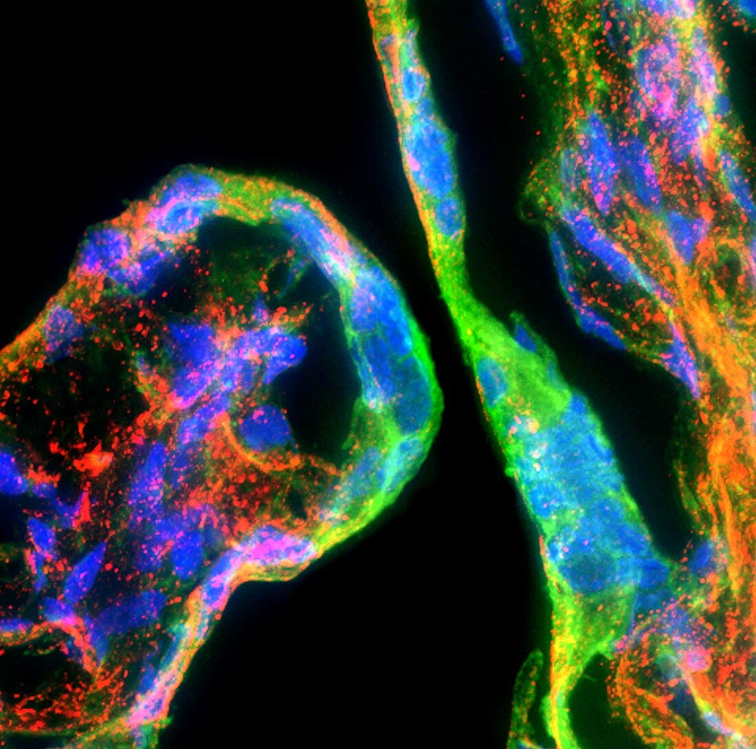
| Formation of placenta,fluorescence deconvolution micrograph. Fluorescent dyes have been used to highlight cellular structures and proteins: cell nuclei (blue),actin (green),PLAC1 protein (red). Actin is a protein that is a major part of a cell's cytoskeleton. PLAC1 (placenta-specific 1) is a protein specific to the placenta. This image shows nuclei of syncitiotrophoblasts on placental villi. The placental villi are structures on the surface of the trophoblast layer of the blastocyst,which is the structure formed after fertilisation. The trophoblast layer will eventually form the placenta,which will support an embryo (not seen) | |
| Lizenzart: | Lizenzpflichtig |
| Credit: | Science Photo Library / R. BICK, B. POINDEXTER, UT MEDICAL SCHOOL |
| Bildgröße: | 3300 px × 3266 px |
| Modell-Rechte: | nicht erforderlich |
| Eigentums-Rechte: | nicht erforderlich |
| Restrictions: | - |
Preise für dieses Bild ab 15 €
Universitäten & Organisationen
(Informationsmaterial Digital, Informationsmaterial Print, Lehrmaterial Digital etc.)
ab 15 €
Redaktionell
(Bücher, Bücher: Sach- und Fachliteratur, Digitale Medien (redaktionell) etc.)
ab 30 €
Werbung
(Anzeigen, Aussenwerbung, Digitale Medien, Fernsehwerbung, Karten, Werbemittel, Zeitschriften etc.)
ab 55 €
Handelsprodukte
(bedruckte Textilie, Kalender, Postkarte, Grußkarte, Verpackung etc.)
ab 75 €
Pauschalpreise
Rechtepakete für die unbeschränkte Bildnutzung in Print oder Online
ab 495 €
Keywords
- Aktin,
- Anatomie,
- anatomisch,
- Biochemie,
- biochemisch,
- dapi,
- Eiweiß,
- Embryologie,
- Entwicklung,
- Entwicklungsbiologie,
- Farbstoff,
- Fluoreszenz,
- Fluoreszenzentfaltung,
- fluoreszierend,
- Gewebe,
- Histologie,
- histologisch,
- Lichtmikroskop,
- Marker,
- menschlicher Körper,
- Mikrofotografie,
- trophoblast,
- Zellbilogie,
- Zelle,
- Zellen,
- Zellkern,
- Zytologie