Embryo spinal cord,light micrograph
Bildnummer 11875437
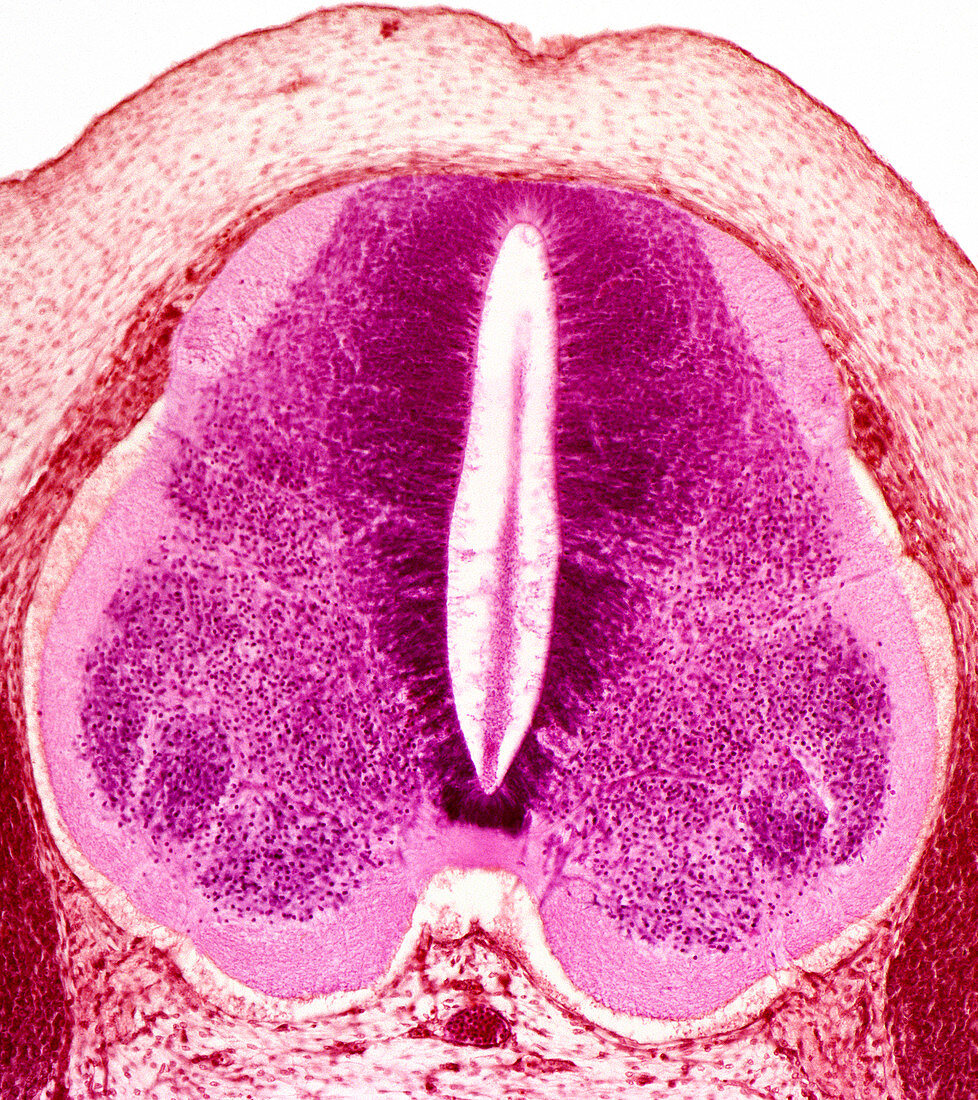
| Embryo spinal cord. Coloured light micrograph of a section through the spinal cord of a 6-week-old human embryo. This spinal cord precursor is known as the neural tube. The central canal (white) is surrounded by structures that will form the grey matter of the central nervous system. At top (dorsal side,facing the back) is the roof plate. Below this is the sulcus limitans (dark pink fibres) and the alar plate (mid-pink,upper centre region). The lower centre region (also mid-pink) is the basal plate. At bottom (ventral side,facing the chest) is the floor plate. The pale pink region around these features will become white matter. The red regions are somites,structures that form skin,muscle and vertebrae (backbones) | |
| Lizenzart: | Lizenzpflichtig |
| Credit: | Science Photo Library / Gschmeissner, Steve |
| Bildgröße: | 3707 px × 4170 px |
| Modell-Rechte: | nicht erforderlich |
| Eigentums-Rechte: | nicht erforderlich |
| Restrictions: | - |
Preise für dieses Bild ab 15 €
Universitäten & Organisationen
(Informationsmaterial Digital, Informationsmaterial Print, Lehrmaterial Digital etc.)
ab 15 €
Redaktionell
(Bücher, Bücher: Sach- und Fachliteratur, Digitale Medien (redaktionell) etc.)
ab 30 €
Werbung
(Anzeigen, Aussenwerbung, Digitale Medien, Fernsehwerbung, Karten, Werbemittel, Zeitschriften etc.)
ab 55 €
Handelsprodukte
(bedruckte Textilie, Kalender, Postkarte, Grußkarte, Verpackung etc.)
ab 75 €
Pauschalpreise
Rechtepakete für die unbeschränkte Bildnutzung in Print oder Online
ab 495 €
Keywords
- Anatomie,
- anatomisch,
- Bildung,
- Biologie,
- biologisch,
- diagonal,
- dorsal,
- eingefärbt,
- Embryo,
- Embryologie,
- embryonale Entwicklung,
- Entwicklung,
- Entwicklungsbiologie,
- farbig,
- Formation,
- fötal,
- Fötus,
- gefärbt,
- Gewebe,
- Grau,
- Histologie,
- histologisch,
- Lichtmikroskop,
- lichtmikroskopische Aufnahme,
- Mensch,
- Menschen,
- menschlicher Körper,
- Mikroskopie,
- Person,
- Querschnitt,
- Rückenmark,
- Scheibe,
- Schichten,
- Sektion,
- Somiten,
- weiße Substanz