5 week embryo,3-D ultrasound scan
Bildnummer 11875363
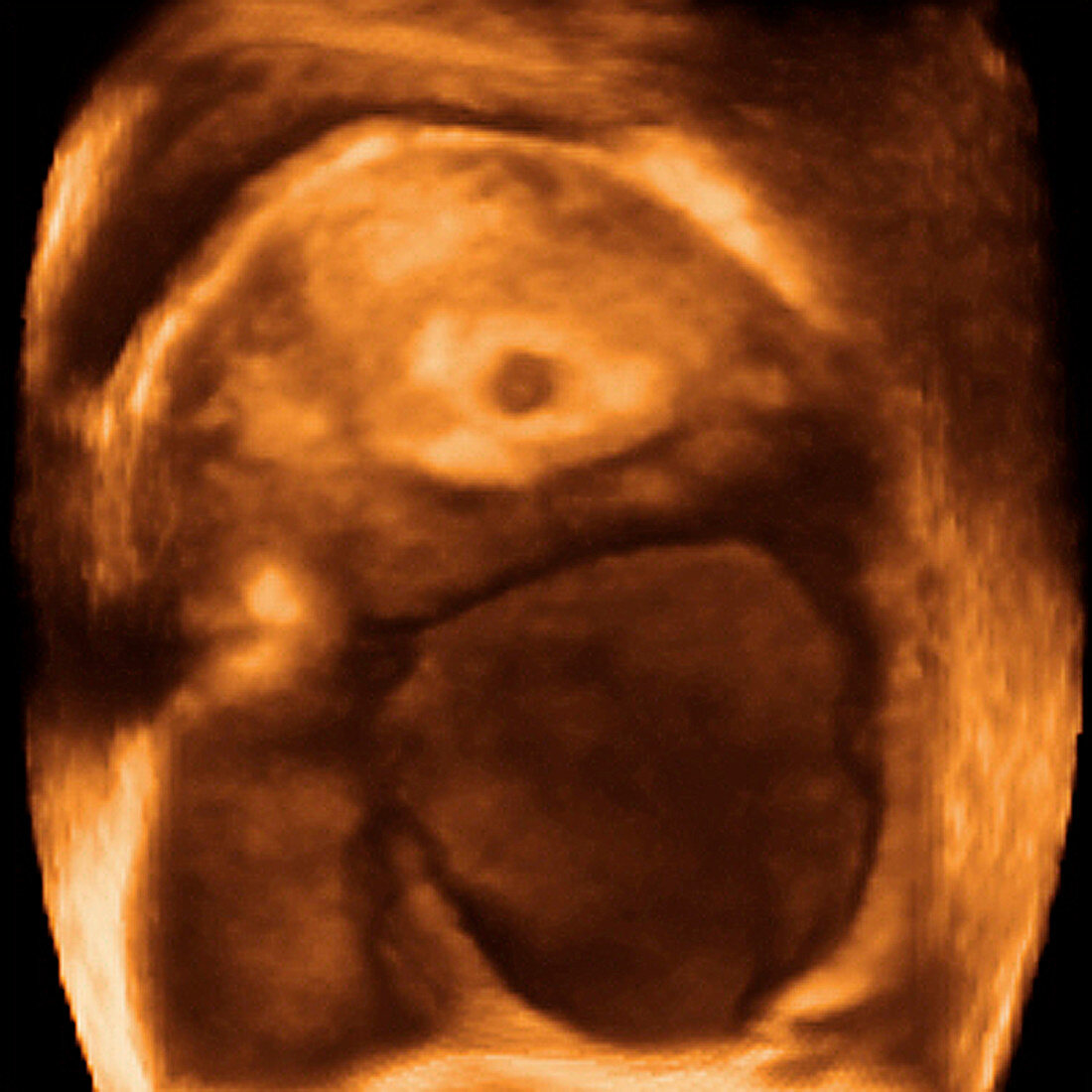
| Embryo. Transverse coloured 3-D ultrasound scan through the abdomen of a pregnant women. The uterus (womb) is at top. Within the uterus is a 5 week embryo (dark circle,upper centre). The dark brown area below the uterus is the pouch of Douglas,part of the peritoneal cavity that lies between the rectum and the back of the uterus. Ultrasound scanning is a diagnostic technique that sends high-frequency sound waves into the body via a transducer. The returning echoes are recorded and used to build an image of an internal structure. Foetal ultrasound scanning is routine during pregnancy. 3-D scanning uses computer technology to return more detailed images than conventional 2-D scans | |
| Lizenzart: | Lizenzpflichtig |
| Credit: | Science Photo Library / Layyous, Dr. Najeeb |
| Bildgröße: | 2555 px × 2555 px |
| Modell-Rechte: | nicht erforderlich |
| Eigentums-Rechte: | nicht erforderlich |
| Restrictions: | - |
Preise für dieses Bild ab 15 €
Universitäten & Organisationen
(Informationsmaterial Digital, Informationsmaterial Print, Lehrmaterial Digital etc.)
ab 15 €
Redaktionell
(Bücher, Bücher: Sach- und Fachliteratur, Digitale Medien (redaktionell) etc.)
ab 30 €
Werbung
(Anzeigen, Aussenwerbung, Digitale Medien, Fernsehwerbung, Karten, Werbemittel, Zeitschriften etc.)
ab 55 €
Handelsprodukte
(bedruckte Textilie, Kalender, Postkarte, Grußkarte, Verpackung etc.)
ab 75 €
Pauschalpreise
Rechtepakete für die unbeschränkte Bildnutzung in Print oder Online
ab 495 €
Keywords
- 3-d,
- 3D,
- Abdomen,
- Baby,
- Diagnose,
- Dreidimensional,
- Embryo,
- Entwicklung,
- farbig,
- Gebärmutter,
- Geburtshilfe,
- geburtshilflich,
- gefärbt,
- gesund,
- Gesundheitswesen,
- Hohlraum,
- Mensch,
- menschlicher Körper,
- normal,
- Orange,
- Reproduktion,
- Scan,
- Scanner,
- Schallwellen,
- schwanger,
- Schwangerschaft,
- Sonographie,
- Ultraschall,
- Ultraschalluntersuchung,
- Uterus