Embryo at 4 weeks,3-D ultrasound scan
Bildnummer 11875281
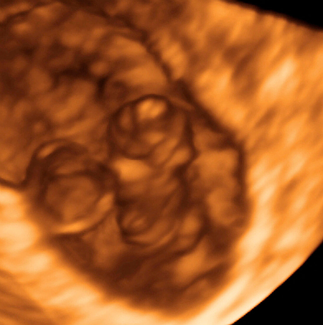
| Embryo and yolk sac. Coloured 3-D ultrasound scan of a 30-day-old embryo (centre) and its yolk sac (left) attached to the wall of the uterus. The yolk sac provides nourishment for the embryo until the umbilical cord is fully functional. The yolk sac also produces blood cells,until the embryo's liver starts to make them at around seven weeks. Ultrasound scanning is a diagnostic technique that sends high-frequency sound waves into the body via a transducer. The returning echoes are recorded and used to build an image of an internal structure. 3-D scanning uses computer technology to return more detailed images than conventional 2-D scans | |
| Lizenzart: | Lizenzpflichtig |
| Credit: | Science Photo Library / Layyous, Dr. Najeeb |
| Bildgröße: | 3373 px × 3390 px |
| Modell-Rechte: | nicht erforderlich |
| Eigentums-Rechte: | nicht erforderlich |
| Restrictions: | - |
Preise für dieses Bild ab 15 €
Universitäten & Organisationen
(Informationsmaterial Digital, Informationsmaterial Print, Lehrmaterial Digital etc.)
ab 15 €
Redaktionell
(Bücher, Bücher: Sach- und Fachliteratur, Digitale Medien (redaktionell) etc.)
ab 30 €
Werbung
(Anzeigen, Aussenwerbung, Digitale Medien, Fernsehwerbung, Karten, Werbemittel, Zeitschriften etc.)
ab 55 €
Handelsprodukte
(bedruckte Textilie, Kalender, Postkarte, Grußkarte, Verpackung etc.)
ab 75 €
Pauschalpreise
Rechtepakete für die unbeschränkte Bildnutzung in Print oder Online
ab 495 €