Four-cell embryo
Bildnummer 11875144
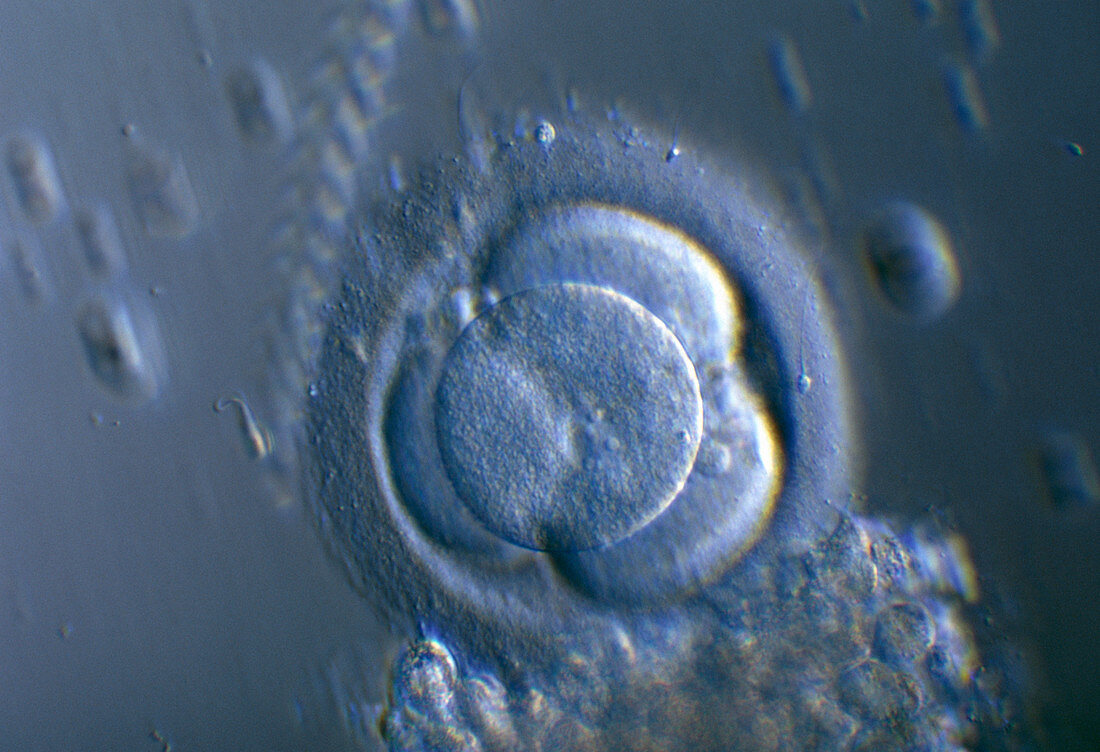
| Four-cell embryo. Light micrograph of a 4-cell human embryo. The cells,or blastomeres,result from two divisions of the fertilized egg. This stage is normally reached around 40 hours after fertilisation. The cells are surrounded by the zona pellucida layer. Outside this at lower right are cumulus corona cells (small circles),the remains of the corona radiata which supported and nourished the egg while it was in the ovary. Photographed at the Hospital Cochin,Paris,France. Magnification unknown | |
| Lizenzart: | Lizenzpflichtig |
| Credit: | Science Photo Library / Goetgheluck, Pascal |
| Bildgröße: | 5184 px × 3543 px |
| Modell-Rechte: | nicht erforderlich |
| Eigentums-Rechte: | nicht erforderlich |
| Restrictions: | - |
Preise für dieses Bild ab 15 €
Universitäten & Organisationen
(Informationsmaterial Digital, Informationsmaterial Print, Lehrmaterial Digital etc.)
ab 15 €
Redaktionell
(Bücher, Bücher: Sach- und Fachliteratur, Digitale Medien (redaktionell) etc.)
ab 30 €
Werbung
(Anzeigen, Aussenwerbung, Digitale Medien, Fernsehwerbung, Karten, Werbemittel, Zeitschriften etc.)
ab 55 €
Handelsprodukte
(bedruckte Textilie, Kalender, Postkarte, Grußkarte, Verpackung etc.)
ab 75 €
Pauschalpreise
Rechtepakete für die unbeschränkte Bildnutzung in Print oder Online
ab 495 €