SEM of a hatched blastocyst 6 days old
Bildnummer 11875068
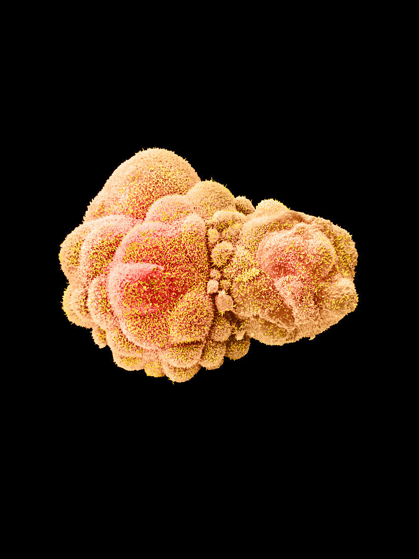
| Hatched blastocyst embryo. Coloured scanning electron micrograph (SEM) of a human embryo at the blastocyst stage,six days after fertilisation. It has fully hatched from the zona pellucida (not seen),a protein shell that originally surrounded the unfertilised egg. The blastocyst is a hollow ball of cells with a fluid centre. Each cell is called a blastomere. Most of these embryo cells will form the placenta and membranes around the embryo,and only a small group (the inner mass) form the embryo proper. At this stage the blastocyst is in the uterus (womb) and is ready to implant on the endometrial wall of the womb. Magnification: x520 at 6x7cm size. Magnification;x480 at original 6x7cm size | |
| Lizenzart: | Lizenzpflichtig |
| Credit: | Science Photo Library / Nikas, Dr. Yorgos |
| Bildgröße: | 2400 px × 3200 px |
| Modell-Rechte: | nicht erforderlich |
| Eigentums-Rechte: | nicht erforderlich |
| Restrictions: | - |
Preise für dieses Bild ab 15 €
Universitäten & Organisationen
(Informationsmaterial Digital, Informationsmaterial Print, Lehrmaterial Digital etc.)
ab 15 €
Redaktionell
(Bücher, Bücher: Sach- und Fachliteratur, Digitale Medien (redaktionell) etc.)
ab 30 €
Werbung
(Anzeigen, Aussenwerbung, Digitale Medien, Fernsehwerbung, Karten, Werbemittel, Zeitschriften etc.)
ab 55 €
Handelsprodukte
(bedruckte Textilie, Kalender, Postkarte, Grußkarte, Verpackung etc.)
ab 75 €
Pauschalpreise
Rechtepakete für die unbeschränkte Bildnutzung in Print oder Online
ab 495 €