Coloured SEM of human embryo at the 2-cell stage
Bildnummer 11875057
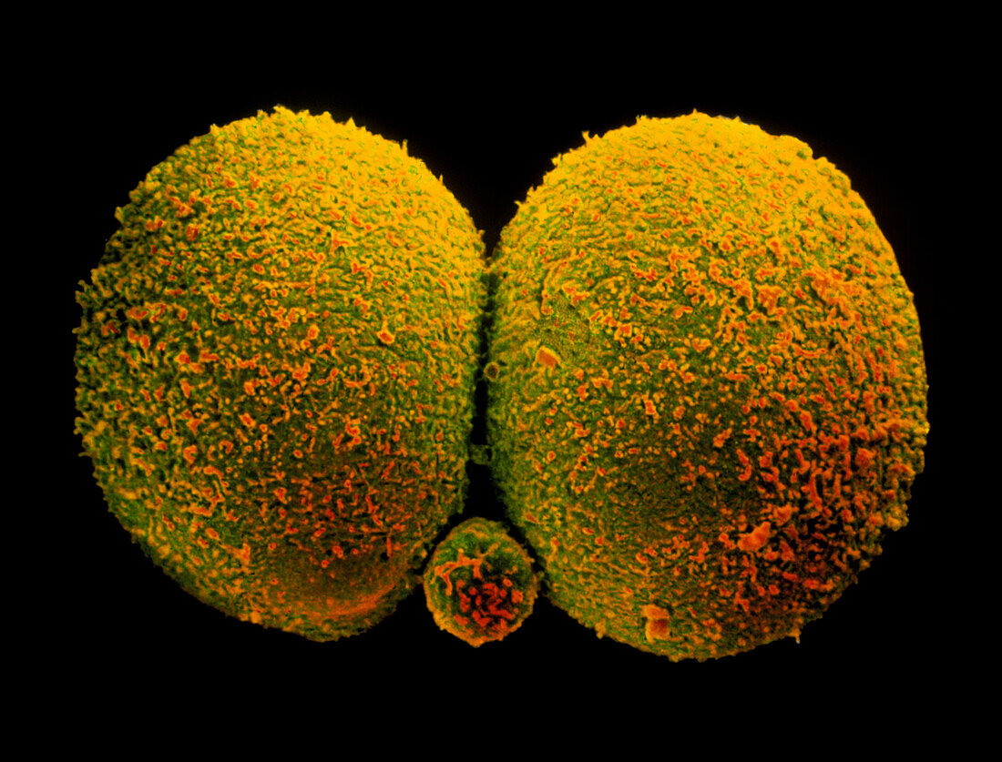
| Two-cell embryo. Coloured scanning electron micrograph (SEM) of a human embryo at the two cell stage,about 30 hours after fertilisation. Still to form into a morula,a cluster of cells,each large rounded cell here is called a blastomere. The smaller spherical structure (at lower centre) is a polar body which will degenerate. The surface of each cell is covered in microvilli. This embryo has only undergone one cell division. The cells will continue to divide and eventually will transform into a human foetus composed of millions of cells. At this two-celled stage,the embryo has not yet implanted in the uterus (womb). Magnification: x930 at 6x7cm size. Magnification; at 8x10 inch size | |
| Lizenzart: | Lizenzpflichtig |
| Credit: | Science Photo Library / Nikas, Dr. Yorgos |
| Bildgröße: | 3543 px × 2696 px |
| Modell-Rechte: | nicht erforderlich |
| Eigentums-Rechte: | nicht erforderlich |
| Restrictions: | - |
Preise für dieses Bild ab 15 €
Universitäten & Organisationen
(Informationsmaterial Digital, Informationsmaterial Print, Lehrmaterial Digital etc.)
ab 15 €
Redaktionell
(Bücher, Bücher: Sach- und Fachliteratur, Digitale Medien (redaktionell) etc.)
ab 30 €
Werbung
(Anzeigen, Aussenwerbung, Digitale Medien, Fernsehwerbung, Karten, Werbemittel, Zeitschriften etc.)
ab 55 €
Handelsprodukte
(bedruckte Textilie, Kalender, Postkarte, Grußkarte, Verpackung etc.)
ab 75 €
Pauschalpreise
Rechtepakete für die unbeschränkte Bildnutzung in Print oder Online
ab 495 €