Coloured SEM of an embryo at the stage of morula
Bildnummer 11874990
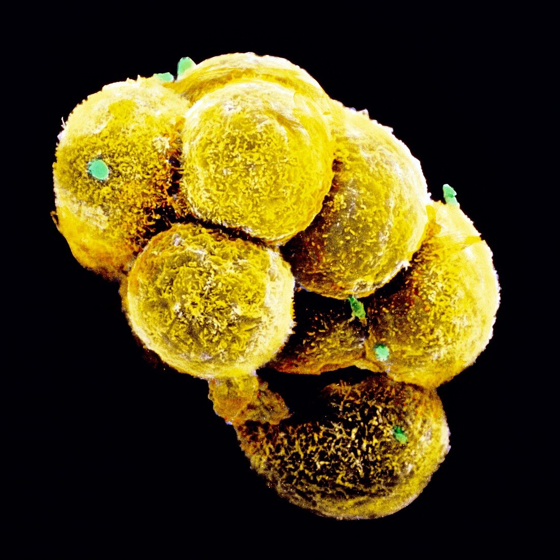
| Embryo development. Coloured scanning electron micrograph of an embryo at the early stage known as the morula. The egg reaches this phase about 4 days after fertilisation after a series of mitotic divisions. At this stage about 12-16 cells are present and are surrounded by a thin glycoprotein layer,the zona pellucida,which was here removed. The inner cells of the morula will give rise to the tissues of the embryo while the outer cells,covered here by microvilli (tiny yellow ridges),will form the placenta. The morula will implant into the uterus six days after fertilisation. Magnification: x1350 at 6x7cm size | |
| Lizenzart: | Lizenzpflichtig |
| Credit: | Science Photo Library / PROFESSORS P.M. MOTTA & J. VAN BLERKOM |
| Bildgröße: | 4228 px × 4228 px |
| Modell-Rechte: | nicht erforderlich |
| Eigentums-Rechte: | nicht erforderlich |
| Restrictions: | - |
Preise für dieses Bild ab 15 €
Universitäten & Organisationen
(Informationsmaterial Digital, Informationsmaterial Print, Lehrmaterial Digital etc.)
ab 15 €
Redaktionell
(Bücher, Bücher: Sach- und Fachliteratur, Digitale Medien (redaktionell) etc.)
ab 30 €
Werbung
(Anzeigen, Aussenwerbung, Digitale Medien, Fernsehwerbung, Karten, Werbemittel, Zeitschriften etc.)
ab 55 €
Handelsprodukte
(bedruckte Textilie, Kalender, Postkarte, Grußkarte, Verpackung etc.)
ab 75 €
Pauschalpreise
Rechtepakete für die unbeschränkte Bildnutzung in Print oder Online
ab 495 €