Human embryo showing eye
Bildnummer 11874840
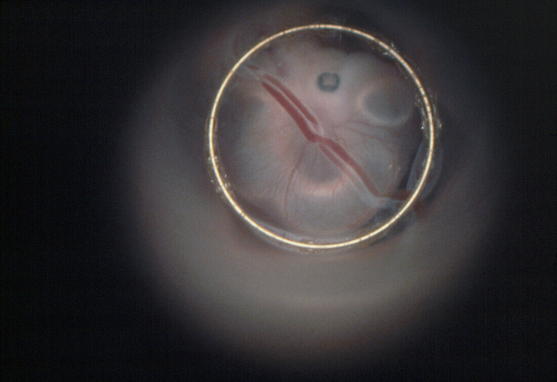
| Endoscopic image of the human embryo in vivo after 4 weeks,showing the retina of the eye (dark spot)- after another week the lens and cornea will form in front of it. To the right is the developing brain. The blood vessels (running diagonally,left to right) connect the embryo to its yolk sac,which continues to supply it with nourishment until the sixth week of development. The endoscope,a series of lenses and a fibre-optic light source,is inserted through the cervix and manipulated until a "window" into the amniotic sac is found (an area where the amniotic wall is thinner) | |
| Lizenzart: | Lizenzpflichtig |
| Credit: | Science Photo Library / Tsiaras, Alexander |
| Bildgröße: | 3614 px × 2480 px |
| Modell-Rechte: | nicht erforderlich |
| Eigentums-Rechte: | nicht erforderlich |
| Restrictions: |
|
Preise für dieses Bild ab 15 €
Universitäten & Organisationen
(Informationsmaterial Digital, Informationsmaterial Print, Lehrmaterial Digital etc.)
ab 15 €
Redaktionell
(Bücher, Bücher: Sach- und Fachliteratur, Digitale Medien (redaktionell) etc.)
ab 30 €
Werbung
(Anzeigen, Aussenwerbung, Digitale Medien, Fernsehwerbung, Karten, Werbemittel, Zeitschriften etc.)
ab 55 €
Handelsprodukte
(bedruckte Textilie, Kalender, Postkarte, Grußkarte, Verpackung etc.)
ab 75 €
Pauschalpreise
Rechtepakete für die unbeschränkte Bildnutzung in Print oder Online
ab 495 €