Immunofluorescent micrograph of sea urchin mitosis
Bildnummer 11874813
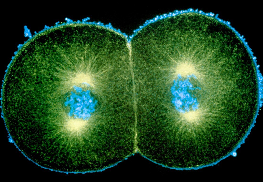
| Mitosis. Immunofluorescence micrograph of a newly fertilized sea urchin embryo dividing into two cells. Each of the daughter cells is about to divide again,and is currently in the prophase of mitosis (division of the nucleus). The chromosomes (blue) have condensed and are beginning to align in the middle of a spindle of fibrous microtubules (green) that will pull them apart into two identi- cal sets. The bright green bodies are the poles of the spindles,or centrioles. This picture was made by treating the embryo with fluorescent anti- bodies that bind to certain proteins in the cells. A laser-scanning light microscope was used to cap- ture the image. Magnification unknown | |
| Lizenzart: | Lizenzpflichtig |
| Credit: | Science Photo Library / PROF. G. SCHATTEN |
| Bildgröße: | 5073 px × 3521 px |
| Modell-Rechte: | nicht erforderlich |
| Eigentums-Rechte: | nicht erforderlich |
| Restrictions: | - |
Preise für dieses Bild ab 15 €
Universitäten & Organisationen
(Informationsmaterial Digital, Informationsmaterial Print, Lehrmaterial Digital etc.)
ab 15 €
Redaktionell
(Bücher, Bücher: Sach- und Fachliteratur, Digitale Medien (redaktionell) etc.)
ab 30 €
Werbung
(Anzeigen, Aussenwerbung, Digitale Medien, Fernsehwerbung, Karten, Werbemittel, Zeitschriften etc.)
ab 55 €
Handelsprodukte
(bedruckte Textilie, Kalender, Postkarte, Grußkarte, Verpackung etc.)
ab 75 €
Pauschalpreise
Rechtepakete für die unbeschränkte Bildnutzung in Print oder Online
ab 495 €