Coloured SEM of folds of the human fallopian tube
Bildnummer 11873868
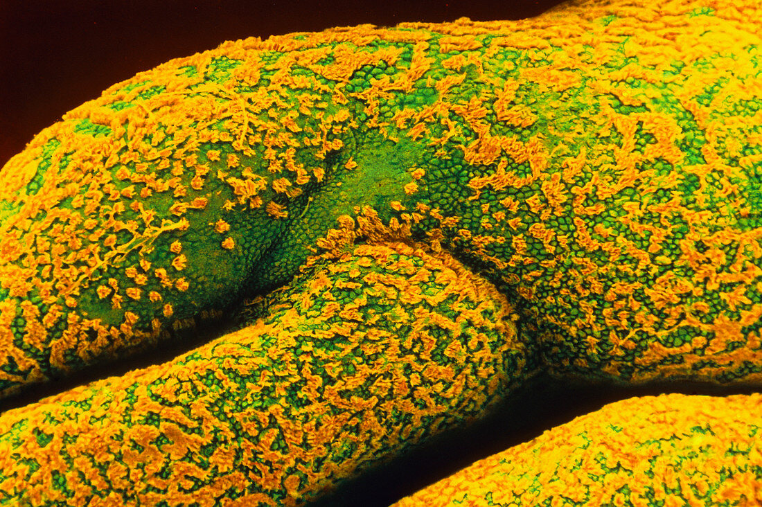
| Fallopian tube surface. Coloured scanning electron micrograph (SEM) of inner surface folds of a human fallopian tube or oviduct. Two fallopian tubes enable eggs from the ovaries of the female to travel to the uterus (womb). Sperm have entered this fallopian tube,seen at upper left with long yellow tails. The surface of the fallopian tube is lined with columnar epithelial cells which here resemble green "scales". There are two types of epithelial cells: ciliated cells with hair-like cilia (yellow tufts); and secretory cells without cilia. Cilia beat rhythmically in the direction of the uterus aiding transport of the egg to the womb. Magnification: x180 at 5x7cm size. x630 at 8x10 | |
| Lizenzart: | Lizenzpflichtig |
| Credit: | Science Photo Library / Nikas, Dr. Yorgos |
| Bildgröße: | 5322 px × 3543 px |
| Modell-Rechte: | nicht erforderlich |
| Eigentums-Rechte: | nicht erforderlich |
| Restrictions: | - |
Preise für dieses Bild ab 15 €
Universitäten & Organisationen
(Informationsmaterial Digital, Informationsmaterial Print, Lehrmaterial Digital etc.)
ab 15 €
Redaktionell
(Bücher, Bücher: Sach- und Fachliteratur, Digitale Medien (redaktionell) etc.)
ab 30 €
Werbung
(Anzeigen, Aussenwerbung, Digitale Medien, Fernsehwerbung, Karten, Werbemittel, Zeitschriften etc.)
ab 55 €
Handelsprodukte
(bedruckte Textilie, Kalender, Postkarte, Grußkarte, Verpackung etc.)
ab 75 €
Pauschalpreise
Rechtepakete für die unbeschränkte Bildnutzung in Print oder Online
ab 495 €