Coloured SEM of section through uterus endometrium
Bildnummer 11873842
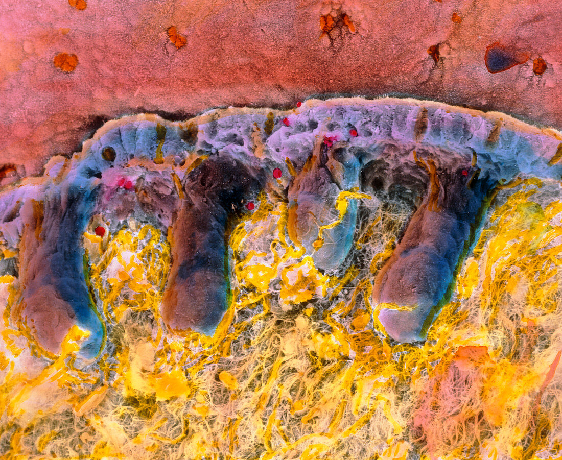
| Uterus endometrium. Coloured Scanning Electron Micrograph (SEM) of a section through the inner wall (endometrium) of the uterus. At top (pink) is the surface of the endometrium containing openings to tubular uterine glands. Epithelial cells making up this wall run left to right (upper centre,blue). Beneath the epithelium are seen the tubular bodies of uterine glands (brown). The glands are embedded in abundant collagen and reticular fibres (yellow) of stroma connective tissue. After ovulation the glands become active and secrete substances to nourish the fertilised egg. Magnification: x440 at 6x7cm size. x570 at 4x5ins | |
| Lizenzart: | Lizenzpflichtig |
| Credit: | Science Photo Library / PROFESSOR P.M. MOTTA & E. VIZZA |
| Bildgröße: | 4724 px × 3869 px |
| Modell-Rechte: | nicht erforderlich |
| Eigentums-Rechte: | nicht erforderlich |
| Restrictions: | - |
Preise für dieses Bild ab 15 €
Universitäten & Organisationen
(Informationsmaterial Digital, Informationsmaterial Print, Lehrmaterial Digital etc.)
ab 15 €
Redaktionell
(Bücher, Bücher: Sach- und Fachliteratur, Digitale Medien (redaktionell) etc.)
ab 30 €
Werbung
(Anzeigen, Aussenwerbung, Digitale Medien, Fernsehwerbung, Karten, Werbemittel, Zeitschriften etc.)
ab 55 €
Handelsprodukte
(bedruckte Textilie, Kalender, Postkarte, Grußkarte, Verpackung etc.)
ab 75 €
Pauschalpreise
Rechtepakete für die unbeschränkte Bildnutzung in Print oder Online
ab 495 €