Coloured SEM of a seminiferous tubule
Bildnummer 11873685
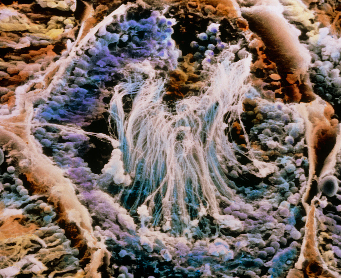
| Testis. Coloured scanning electron micrograph showing a cross section of a seminiferous tubule within a testis. This is the site of production and maturation of sperm. The tubule is surrounded by a basal lamina (pale orange) which separates it from other seminiferous tubules. Round undifferentiated cells (blue-purple) line the tubule wall; they will halve their chromosome number and become mature spermatozoa (centre with tails) in a process which takes about two months. Mature sperm then migrate from the tubule to the epididymis where they are stored. Magnification: x590 at 6x7cm size | |
| Lizenzart: | Lizenzpflichtig |
| Credit: | Science Photo Library / PROFESSORS P.M. MOTTA, K.R. PORTER & P.M. ANDREWS |
| Bildgröße: | 5039 px × 4112 px |
| Modell-Rechte: | nicht erforderlich |
| Eigentums-Rechte: | nicht erforderlich |
| Restrictions: | - |
Preise für dieses Bild ab 15 €
Universitäten & Organisationen
(Informationsmaterial Digital, Informationsmaterial Print, Lehrmaterial Digital etc.)
ab 15 €
Redaktionell
(Bücher, Bücher: Sach- und Fachliteratur, Digitale Medien (redaktionell) etc.)
ab 30 €
Werbung
(Anzeigen, Aussenwerbung, Digitale Medien, Fernsehwerbung, Karten, Werbemittel, Zeitschriften etc.)
ab 55 €
Handelsprodukte
(bedruckte Textilie, Kalender, Postkarte, Grußkarte, Verpackung etc.)
ab 75 €
Pauschalpreise
Rechtepakete für die unbeschränkte Bildnutzung in Print oder Online
ab 495 €