3-D CT scan of normal chest showing lungs
Bildnummer 11873547
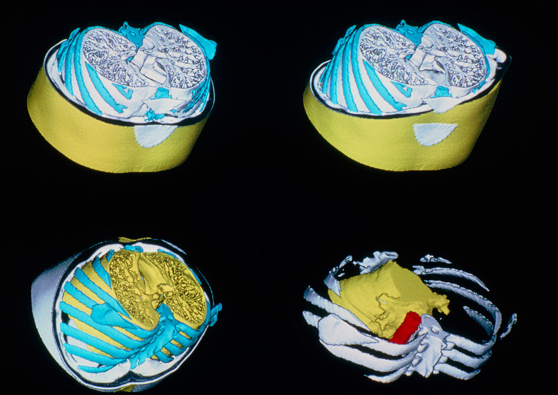
| Four 3-D CT (computed tomography) scans of the chest showing normal lungs: bone is coloured blue,the lungs in yellow & the aorta in red. CT scanning provides clear cross-sectional images of the body. X-ray beams are passed through the body in a circular track; the amount of X-rays absorbed by different tissues is recorded by detectors in the scanner and transformed via a computer into 2- & 3-D images. These images were produced on a Twin CT scanner,a device with a double source/detector system that affords reduced scan times,improved image contrast & resolution & the ability to resolve organ volume | |
| Lizenzart: | Lizenzpflichtig |
| Credit: | Science Photo Library / Plailly, Philippe |
| Bildgröße: | 4883 px × 3459 px |
| Modell-Rechte: | nicht erforderlich |
| Eigentums-Rechte: | nicht erforderlich |
| Restrictions: |
|
Preise für dieses Bild ab 15 €
Universitäten & Organisationen
(Informationsmaterial Digital, Informationsmaterial Print, Lehrmaterial Digital etc.)
ab 15 €
Redaktionell
(Bücher, Bücher: Sach- und Fachliteratur, Digitale Medien (redaktionell) etc.)
ab 30 €
Werbung
(Anzeigen, Aussenwerbung, Digitale Medien, Fernsehwerbung, Karten, Werbemittel, Zeitschriften etc.)
ab 55 €
Handelsprodukte
(bedruckte Textilie, Kalender, Postkarte, Grußkarte, Verpackung etc.)
ab 75 €
Pauschalpreise
Rechtepakete für die unbeschränkte Bildnutzung in Print oder Online
ab 495 €