Trachea and rib cage,3D CT scan
Bildnummer 11873478
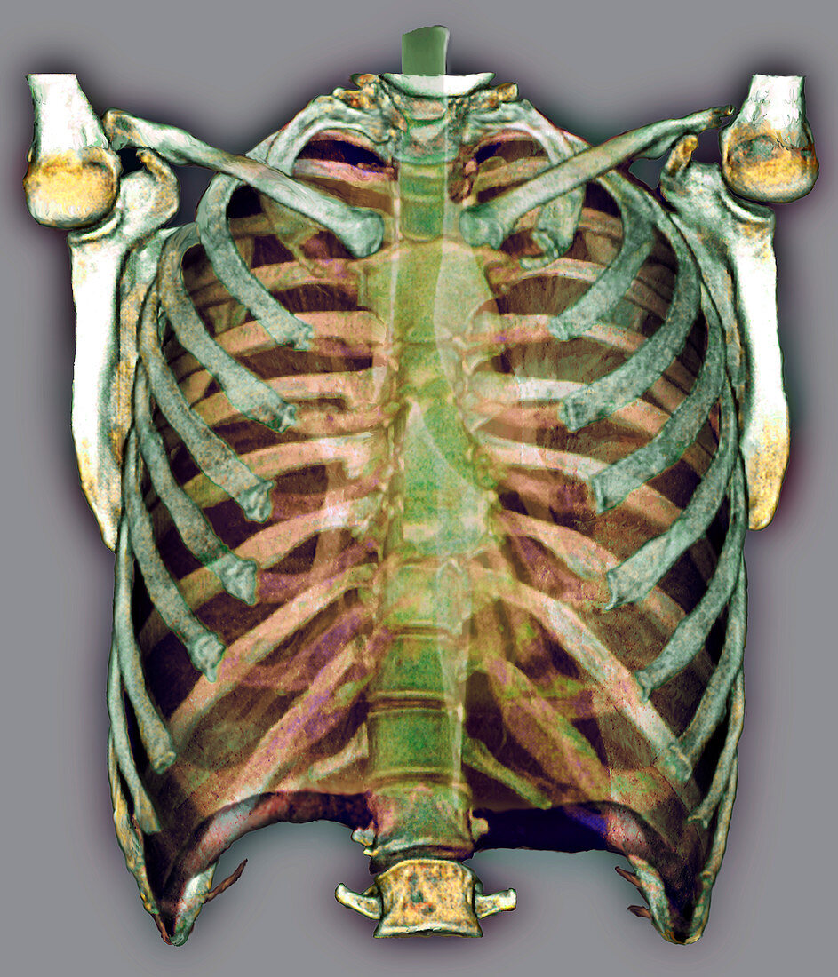
| Trachea and rib cage. Coloured frontal 3D computed tomography (CT) scan of a normal trachea and rib cage. The trachea (windpipe,green) runs from the throat (top centre) down to centre,and then splits into the two bronchi,one for each lung (spaces at left and right). The sternum (breast bone) and the rest of the thoracic bones are shown. Twelve pairs of ribs make up the rib cage enclosing the chest. They are attached at one end to the spine (backbone,down centre). The collar bones (clavicles) lie across the top of the rib cage and attach at one end to the breast bone and at the other ends to the shoulder bones. The shoulder blades (scapulas) and the head of the humerus (upper arm bone) are also seen,forming the shoulder joints | |
| Lizenzart: | Lizenzpflichtig |
| Credit: | Science Photo Library / Zephyr |
| Bildgröße: | 3402 px × 3969 px |
| Modell-Rechte: | nicht erforderlich |
| Eigentums-Rechte: | nicht erforderlich |
| Restrictions: | - |
Preise für dieses Bild ab 15 €
Universitäten & Organisationen
(Informationsmaterial Digital, Informationsmaterial Print, Lehrmaterial Digital etc.)
ab 15 €
Redaktionell
(Bücher, Bücher: Sach- und Fachliteratur, Digitale Medien (redaktionell) etc.)
ab 30 €
Werbung
(Anzeigen, Aussenwerbung, Digitale Medien, Fernsehwerbung, Karten, Werbemittel, Zeitschriften etc.)
ab 55 €
Handelsprodukte
(bedruckte Textilie, Kalender, Postkarte, Grußkarte, Verpackung etc.)
ab 75 €
Pauschalpreise
Rechtepakete für die unbeschränkte Bildnutzung in Print oder Online
ab 495 €
Keywords
- 3-d,
- 3D,
- Anatomie,
- anatomisch,
- anterior,
- Atmungssystem,
- Biologie,
- biologisch,
- Clavicula,
- Computertomographie,
- CT-Scan,
- CT-Scanner,
- Dreidimensional,
- Erwachsene,
- farbig,
- Frontal,
- gefärbt,
- gesund,
- Gesundheitswesen,
- Klinge,
- Knochen,
- Luftröhre,
- menschlicher Körper,
- normal,
- Organ,
- pulmonal,
- Rippen,
- Rückgrat,
- Scanner,
- Schlüsselbeine,
- Schulterblatt,
- Schulterblätter,
- Sternum,
- thorakal,
- Thorax,
- Truhe,
- Wirbelsäule