LM of a longitudinal section of a bronchus
Bildnummer 11873403
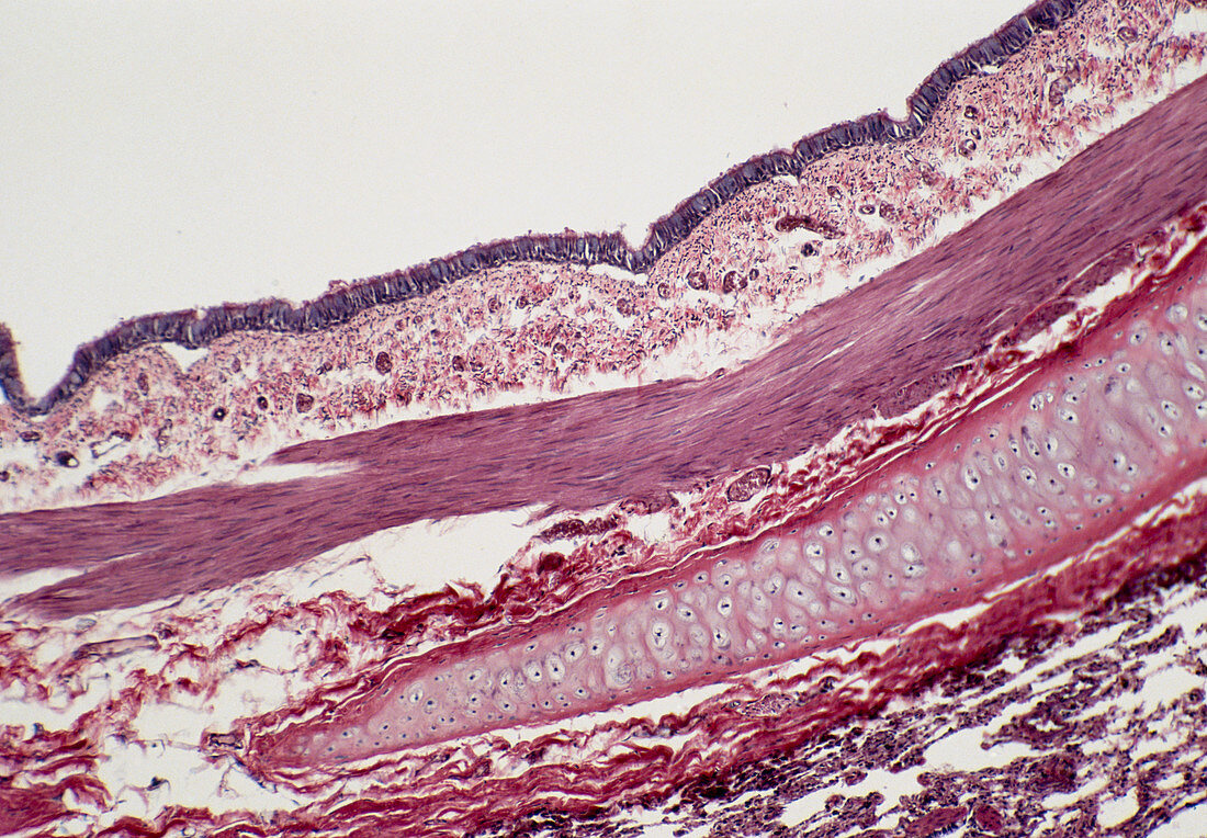
| Light micrograph of a longitudinal section of a bronchus. The epithelium (dark violet at top) is ciliated and goblet cells,which secrete mucus,are occasionally found. It is supported by a thick layer of highly vascular connective tissue known as the lamina propria. This is separated from the submucosa by a layer of smooth muscle (pink and compact). The vacuolated sheath running from centre right to bottom left is a layer formed by hyaline cartilage. Magnification: x25 at 35mm size | |
| Lizenzart: | Lizenzpflichtig |
| Credit: | Science Photo Library / Michler, Astrid & Hans-Frieder |
| Bildgröße: | 5014 px × 3484 px |
| Modell-Rechte: | nicht erforderlich |
| Eigentums-Rechte: | nicht erforderlich |
| Restrictions: | - |
Preise für dieses Bild ab 15 €
Universitäten & Organisationen
(Informationsmaterial Digital, Informationsmaterial Print, Lehrmaterial Digital etc.)
ab 15 €
Redaktionell
(Bücher, Bücher: Sach- und Fachliteratur, Digitale Medien (redaktionell) etc.)
ab 30 €
Werbung
(Anzeigen, Aussenwerbung, Digitale Medien, Fernsehwerbung, Karten, Werbemittel, Zeitschriften etc.)
ab 55 €
Handelsprodukte
(bedruckte Textilie, Kalender, Postkarte, Grußkarte, Verpackung etc.)
ab 75 €
Pauschalpreise
Rechtepakete für die unbeschränkte Bildnutzung in Print oder Online
ab 495 €