LM of a longitudinal section of the trachea
Bildnummer 11873402
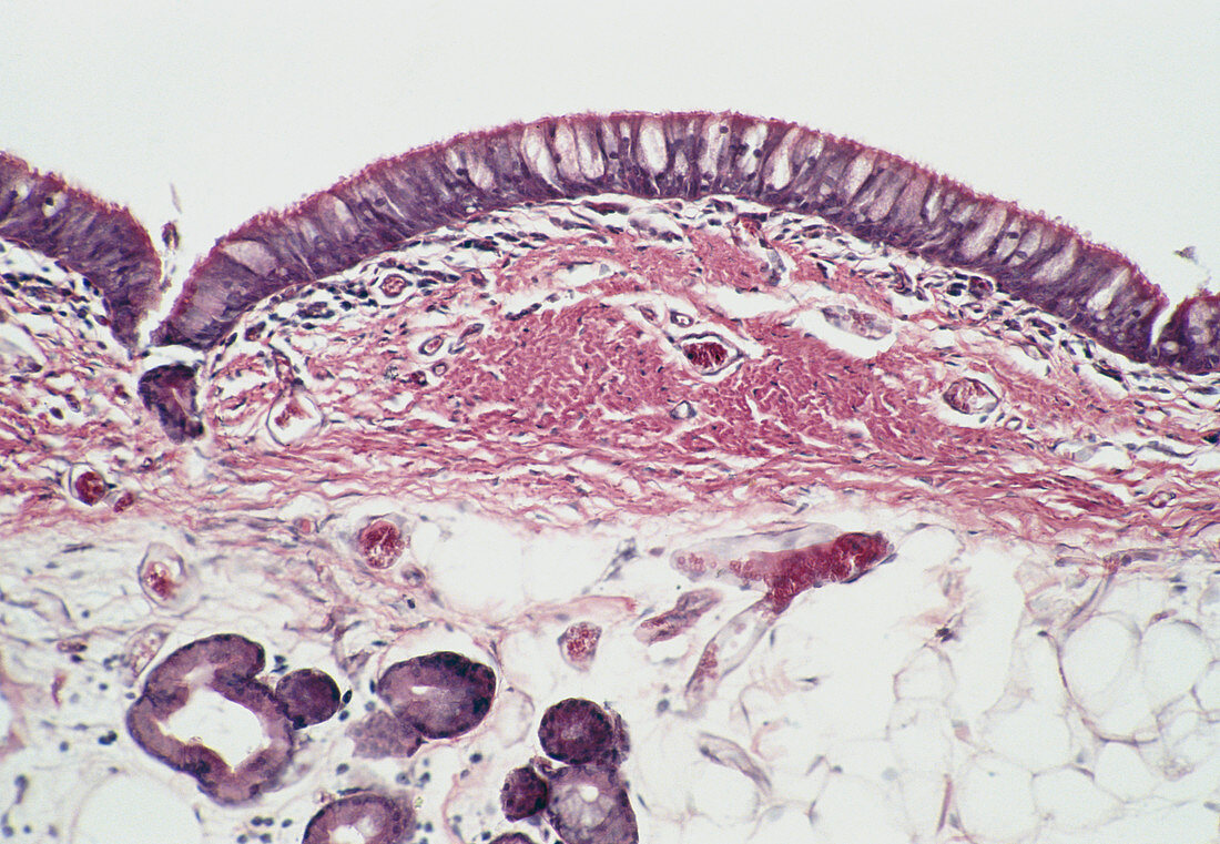
| Light micrograph of a longitudinal section of the trachea with lumen (white) at top. The epithelium (dark violet at top) is tall,pseudostratified and ciliated and contains numerous goblet cells (light violet). The epithelium is supported by a basement membrane which is not clearly visible here. Beneath this membrane there is a layer of loose,highly vascular connective tissue,the lamina propria,which becomes condensed at centre to form a band of fibro-elastic tissue. The loose submucosa (bottom) contains numerous mixed sero-mucous glands. A group of these is visible at bottom left. Magnification: x50 at 35mm size | |
| Lizenzart: | Lizenzpflichtig |
| Credit: | Science Photo Library / Michler, Astrid & Hans-Frieder |
| Bildgröße: | 5195 px × 3592 px |
| Modell-Rechte: | nicht erforderlich |
| Eigentums-Rechte: | nicht erforderlich |
| Restrictions: | - |
Preise für dieses Bild ab 15 €
Universitäten & Organisationen
(Informationsmaterial Digital, Informationsmaterial Print, Lehrmaterial Digital etc.)
ab 15 €
Redaktionell
(Bücher, Bücher: Sach- und Fachliteratur, Digitale Medien (redaktionell) etc.)
ab 30 €
Werbung
(Anzeigen, Aussenwerbung, Digitale Medien, Fernsehwerbung, Karten, Werbemittel, Zeitschriften etc.)
ab 55 €
Handelsprodukte
(bedruckte Textilie, Kalender, Postkarte, Grußkarte, Verpackung etc.)
ab 75 €
Pauschalpreise
Rechtepakete für die unbeschränkte Bildnutzung in Print oder Online
ab 495 €