Heart and lungs,CT scan
Bildnummer 11873376
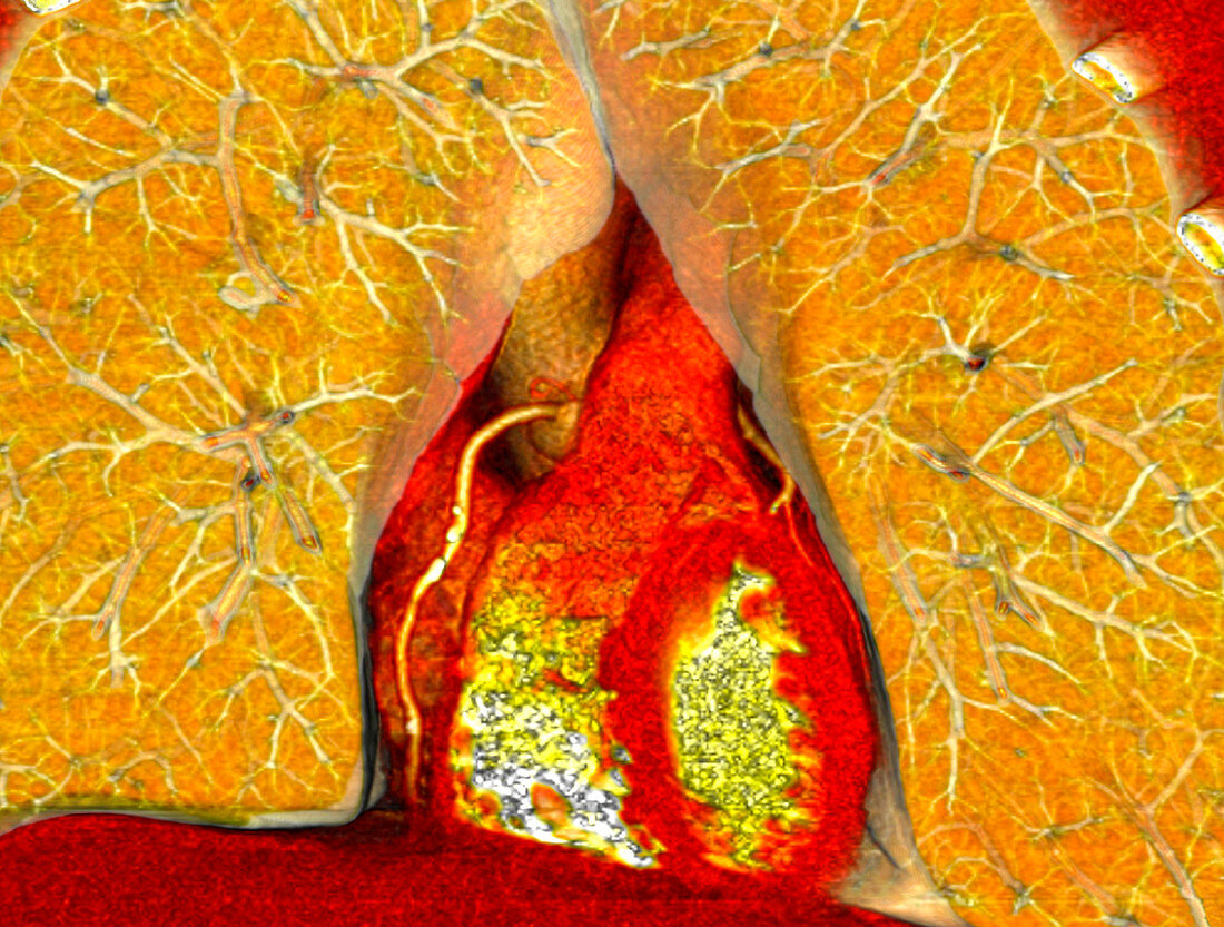
| Heart and lungs,CT scan. Coloured 3-D computed tomography (CT) scan of the heart (lower centre) and lungs. The vena cava,aorta and pulmonary artery are visible emerging from the top of the heart. The internal chambers of the heart are visible (centre). The blood vessels of the lungs are visible to the left and right sides of the heart. The rib cage (top right) surrounds the lungs. This image was produced using a multi-slice CT scanner,which uses a thin X-ray beam to scan around the patient collecting data from different angles to create 'slices' of the body. Osirix medical imaging software was used to create a coloured 3-D dimensional image from the data | |
| Lizenzart: | Lizenzpflichtig |
| Credit: | Science Photo Library / Rosset, Antoine |
| Bildgröße: | 3402 px × 2576 px |
| Modell-Rechte: | nicht erforderlich |
| Eigentums-Rechte: | nicht erforderlich |
| Restrictions: | - |
Preise für dieses Bild ab 15 €
Universitäten & Organisationen
(Informationsmaterial Digital, Informationsmaterial Print, Lehrmaterial Digital etc.)
ab 15 €
Redaktionell
(Bücher, Bücher: Sach- und Fachliteratur, Digitale Medien (redaktionell) etc.)
ab 30 €
Werbung
(Anzeigen, Aussenwerbung, Digitale Medien, Fernsehwerbung, Karten, Werbemittel, Zeitschriften etc.)
ab 55 €
Handelsprodukte
(bedruckte Textilie, Kalender, Postkarte, Grußkarte, Verpackung etc.)
ab 75 €
Pauschalpreise
Rechtepakete für die unbeschränkte Bildnutzung in Print oder Online
ab 495 €
Keywords
- 3-d,
- 3D,
- Anatomie,
- anatomisch,
- Angiogramm,
- Aorta,
- ärztliche Untersuchung,
- Atmung,
- Atmungssystem,
- Blutgefäße,
- Computertomographie,
- CT-Scan,
- Diagnose,
- diagnostische Bildgebung,
- Dreidimensional,
- farbig,
- gefärbt,
- Herz,
- Herz-Kreislauf,
- kardiovaskular,
- Lunge,
- Lungen,
- Lungenarterie,
- Medizin,
- medizinisch,
- medizinische Bildgebung,
- medizinische Visualisierung,
- medizinischer Scan,
- Mensch,
- Menschen,
- menschlicher Körper,
- OsiriX,
- Person,
- Radiographie,
- Radiologie,
- radiologisch,
- Rippen,
- Röntgen,
- Röntgenstrahlen,
- Röntgenstrahlung,
- Scanner