Kidneys and ureters,X-ray
Bildnummer 11873213
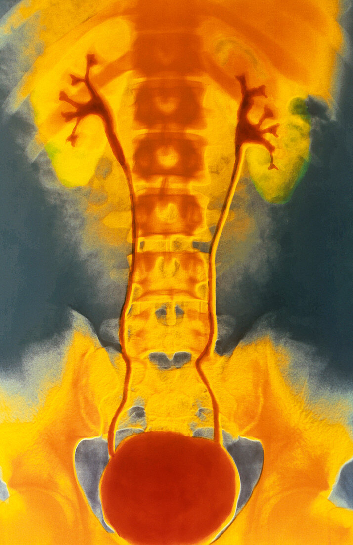
| False-colour X-ray image of the abdominal & pelvic areas of a healthy person,featuring both kidneys (green) & ureters (red - the vessels connecting the kidneys to the bladder,the red circular mass at bottom),outlined using intravenous pyelography (IVP). The technique involves prior intravenous injection of an iodine-based,radio-opaque contrast medium,which is concentrated & excreted by the kidneys. Branches of the renal pelvis (structures of the kidney into which urine is drained),the ureters (25-30 cm long in adults),& the urine-filled bladder are defined. IVP tests kidney function,reveals stones in kidneys or ureters & other malfunctions of the urinary tract | |
| Lizenzart: | Lizenzpflichtig |
| Credit: | Science Photo Library / Pol, Alain / ISM |
| Bildgröße: | 3393 px × 5227 px |
| Modell-Rechte: | nicht erforderlich |
| Eigentums-Rechte: | nicht erforderlich |
| Restrictions: |
|
Preise für dieses Bild ab 15 €
Universitäten & Organisationen
(Informationsmaterial Digital, Informationsmaterial Print, Lehrmaterial Digital etc.)
ab 15 €
Redaktionell
(Bücher, Bücher: Sach- und Fachliteratur, Digitale Medien (redaktionell) etc.)
ab 30 €
Werbung
(Anzeigen, Aussenwerbung, Digitale Medien, Fernsehwerbung, Karten, Werbemittel, Zeitschriften etc.)
ab 55 €
Handelsprodukte
(bedruckte Textilie, Kalender, Postkarte, Grußkarte, Verpackung etc.)
ab 75 €
Pauschalpreise
Rechtepakete für die unbeschränkte Bildnutzung in Print oder Online
ab 495 €